


 全部商品分类


hGlut1 MAb (Cl 20291 (200 ug)

 下载产品说明书 下载SDS
 用小程序,查商品更便捷


 收藏
收藏
 对比
对比 咨询
咨询Immunocytochemistry(8-25 µg/mL)

Met1-Val492
Accession # AAA52571
Scientific Data
 View Larger
Detection of Glut1 in HepG2 Human Cell Line by Flow Cytometry. HepG2 human hepatocellular carcinoma cell line was stained with Mouse Anti-Human Glut1 Monoclonal Antibody (Catalog # MAB1418, filled histogram) or isotype control antibody (Catalog # MAB0041, open histogram), followed by Allophycocyanin-conjugated Anti-Mouse IgG Secondary Antibody (Catalog # F0101B).
 View Larger
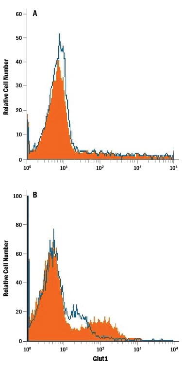
Detection of Glut1 in Jurkat Human Cell Line by Flow Cytometry. Jurkat human acute T cell leukemia cell line either (A) untreated or (B) cultured in nutrient-depleted media was stained with Mouse Anti-Human Glut1 Monoclonal Antibody (Catalog # MAB1418, filled histogram) or isotype control antibody (Catalog # MAB0041, open histogram), followed by Phycoerythrin-conjugated Anti-Mouse IgG Secondary Antibody (Catalog # F0102B).
.") View Larger
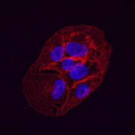
View LargerGlut1 in HepG2 Human Hepatocellular Carcinoma Cell Line. Glut1 was detected in immersion fixed HepG2 human hepatocellular carcinoma cell line using Mouse Anti-Human Glut1 Monoclonal Antibody (Catalog # MAB1418) at 10 µg/mL for 3 hours at room temperature. Cells were stained using the NorthernLights™ 557-conjugated Anti-Mouse IgG Secondary Antibody (red; Catalog # NL007) and counterstained with DAPI(blue). Specific staining was localized to the plasma membrane. View our protocol for Fluorescent ICC Staining of Cells on Coverslips.
Human Glut1 Antibody Summary
Met1-Val492
Accession # AAA52571
Applications
Please Note: Optimal dilutions should be determined by each laboratory for each application. General Protocols are available in the Technical Information section on our website.
Immunocytochemistry(8-25 µg/mL)
Background: Glut1
Glut1 belongs to the facilitative glucose transport protein family that comprises 13 members. It is an integral membrane protein with 12 transmembrane domains and is expressed at variable levels in many tissues including brain endothelial cells, CD8+ T cells, and erythrocytes (1‑4). Glut1 is a major glucose transporter that mediates glucose transport across the mammalian blood‑brain barrier.
- Mueckler, M. et al. 1994, Eur. J. Biochem. 219:713.
- Meuckler, M. et al. 1985, Science 229:941.
- Jones, K.S. et al. 2006, J. Virol. 8291.
- Takenouchi, N. et al. 2007, J. Virol. 1506.
- Kinet, S. et al. 2007, Retrovirology 4:31.
Preparation and Storage
- 12 months from date of receipt, -20 to -70 °C as supplied.
- 1 month, 2 to 8 °C under sterile conditions after reconstitution.
- 6 months, -20 to -70 °C under sterile conditions after reconstitution.
参考图片
Detection of Glut1 in Jurkat Human Cell Line by Flow Cytometry. Jurkat human acute T cell leukemia cell line either (A) untreated or (B) cultured in nutrient-depleted media was stained with Mouse Anti-Human Glut1 Monoclonal Antibody (Catalog # MAB1418, filled histogram) or isotype control antibody (Catalog # MAB0041, open histogram), followed by Phycoerythrin-conjugated Anti-Mouse IgG Secondary Antibody (Catalog # F0102B).
Detection of Glut1 in HepG2 Human Cell Line by Flow Cytometry. HepG2 human hepatocellular carcinoma cell line was stained with Mouse Anti-Human Glut1 Monoclonal Antibody (Catalog # MAB1418, filled histogram) or isotype control antibody (Catalog # MAB0041, open histogram), followed by Allophycocyanin-conjugated Anti-Mouse IgG Secondary Antibody (Catalog # F0101B).
Glut1 in HepG2 Human Hepatocellular Carcinoma Cell Line. Glut1 was detected in immersion fixed HepG2 human hepatocellular carcinoma cell line using Mouse Anti-Human Glut1 Monoclonal Antibody (Catalog # MAB1418) at 10 µg/mL for 3 hours at room temperature. Cells were stained using the NorthernLights™ 557-conjugated Anti-Mouse IgG Secondary Antibody (red; Catalog # NL007) and counterstained with DAPI(blue). Specific staining was localized to the plasma membrane. View our protocol for Fluorescent ICC Staining of Cells on Coverslips.