


 全部商品分类
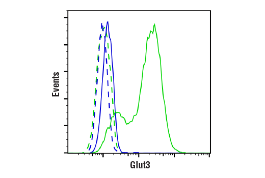


 用小程序,查商品更便捷


Monoclonal antibody is produced by immunizing animals with a synthetic peptide corresponding to residues surrounding Ala472 of human Glut3 protein.
Product Usage Information
| Application | Dilution |
|---|---|
| Western Blotting | 1:1000 |
| Immunohistochemistry (Paraffin) | 1:3200 - 1:12800 |
| Immunofluorescence (Immunocytochemistry) | 1:800 - 1:1600 |
| Flow Cytometry (Fixed/Permeabilized) | 1:50 - 1:200 |
Specificity/Sensitivity
Species Reactivity:
Human
Supplied in 10 mM sodium HEPES (pH 7.5), 150 mM NaCl, 100 µg/ml BSA, 50% glycerol and less than 0.02% sodium azide. Store at –20°C. Do not aliquot the antibody.
For a carrier free (BSA and azide free) version of this product see product #18035.
参考图片
Flow cytometric analysis of OVCAR-4 cells (blue) and SW480 cells (green) using Glut3 (E7M7V) Rabbit mAb (solid lines) or a concentration-matched Rabbit (DA1E) mAb IgG XP® Isotype Control #3900 (dashed lines). Anti-rabbit IgG (H+L), F(ab')2 Fragment (Alexa Fluor® 488 Conjugate) #4412 was used as a secondary antibody.
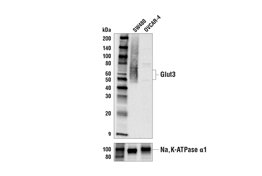
Western blot analysis of membrane/organelle fraction extracts from SW480 and OVCAR-4 cells using Glut3 (E7M7V) Rabbit mAb (upper) and Na,K-ATPase α1 (D4Y7E) Rabbit mAb #23565 (lower). The difference in Glut3 expression levels detected in SW480 and OVCAR-4 extracts is consistent with both RNAseq and proteomic expression profiling data, confirming specificity of the antibody for Glut3.
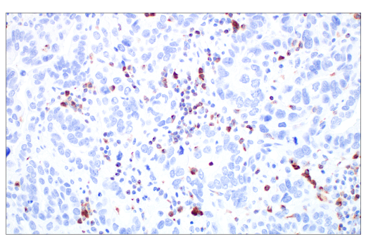
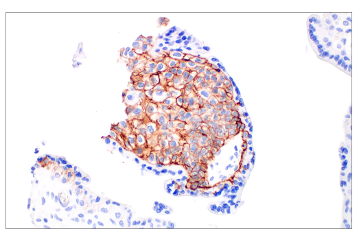
Immunohistochemical analysis of paraffin-embedded human large cell neuroendocrine lung carcinoma using Glut3 (E7M7V) Rabbit mAb.
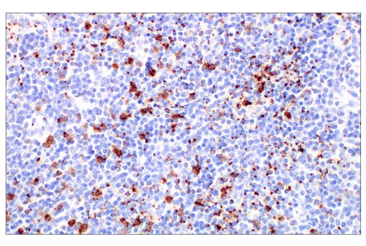
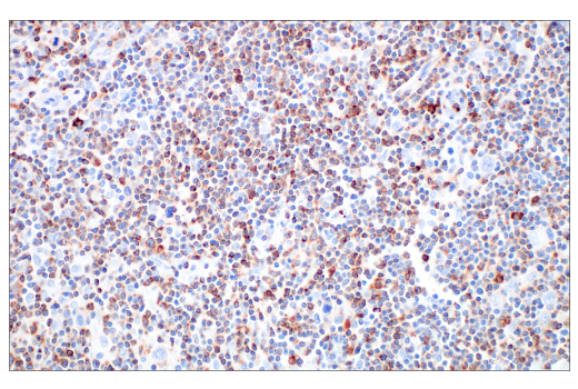
Immunohistochemical analysis of paraffin-embedded human B-cell non-Hodgkin lymphoma using Glut3 (E7M7V) Rabbit mAb.
Immunohistochemical analysis of paraffin-embedded human esophageal adenocarcinoma using Glut3 (E7M7V) Rabbit mAb.
Immunohistochemical analysis of paraffin-embedded human Hodgkin lymphoma using Glut3 (E7M7V) Rabbit mAb.
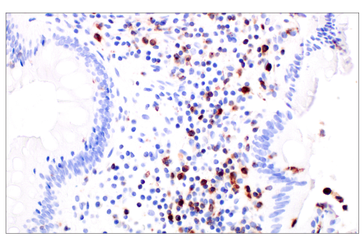
Immunohistochemical analysis of paraffin-embedded human colon adenocarcinoma using Glut3 (E7M7V) Rabbit mAb
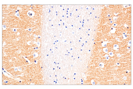
Immunohistochemical analysis of paraffin-embedded normal human brain using Glut3 (E7M7V) Rabbit mAb.
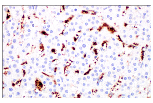
Immunohistochemical analysis of paraffin-embedded normal human adrenal gland using Glut3 (E7M7V) Rabbit mAb.
Immunohistochemical analysis of paraffin-embedded normal human placenta using Glut3 (E7M7V) Rabbit mAb.
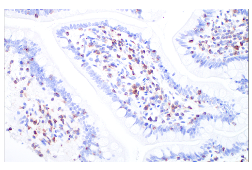
Immunohistochemical analysis of paraffin-embedded normal human small intestine using Glut3 (E7M7V) Rabbit mAb.
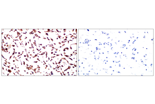
Immunohistochemical analysis of paraffin-embedded normal human testis using Glut3 (E7M7V) Rabbit mAb (left) compared to concentration-matched Rabbit (DA1E) mAb IgG XP® Isotype Control #3900 (right).
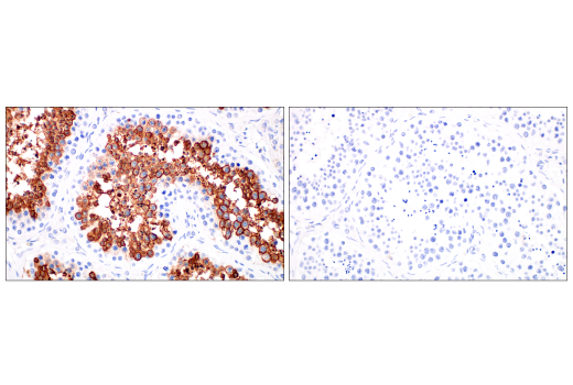
Immunohistochemical analysis of paraffin-embedded SW480 cell pellet (left, positive) or OVCAR-4 cell pellet (right, negative) using Glut3 (E7M7V) Rabbit mAb.
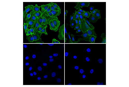
Confocal immunofluorescent analysis of SW480 cells (top, positive) and OVCAR-4 cells (bottom, negative). Cells were fixed with either ice-cold 100% methanol (left) or 4% formaldehyde (right), stained with Glut3 (E7M7V) Rabbit mAb (green), and then mounted in ProLong® Gold Antifade Reagent with DAPI #8961 (blue).