


 全部商品分类
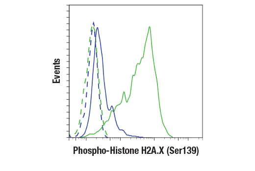


Phospho-Histone H2A.X (Ser139) (20E3) Rabbit mAb


 下载产品说明书 下载COA 下载SDS
 用小程序,查商品更便捷


 收藏
收藏
 对比
对比 咨询
咨询
Monoclonal antibody is produced by immunizing animals with a synthetic phosphopeptide corresponding to residues surrounding Ser139 of human H2A.X.
Product Usage Information
| Application | Dilution |
|---|---|
| Western Blotting | 1:1000 |
| IHC Leica Bond | 1:50 - 1:200 |
| Immunohistochemistry (Paraffin) | 1:240 - 1:960 |
| Immunofluorescence (Immunocytochemistry) | 1:200 - 1:800 |
| Flow Cytometry (Fixed/Permeabilized) | 1:100 - 1:400 |
Specificity/Sensitivity
Species Reactivity:
Human, Mouse, Rat, Monkey
Supplied in 10 mM sodium HEPES (pH 7.5), 150 mM NaCl, 100 µg/ml BSA, 50% glycerol and less than 0.02% sodium azide. Store at –20°C. Do not aliquot the antibody.
For a carrier free (BSA and azide free) version of this product see product #60566.
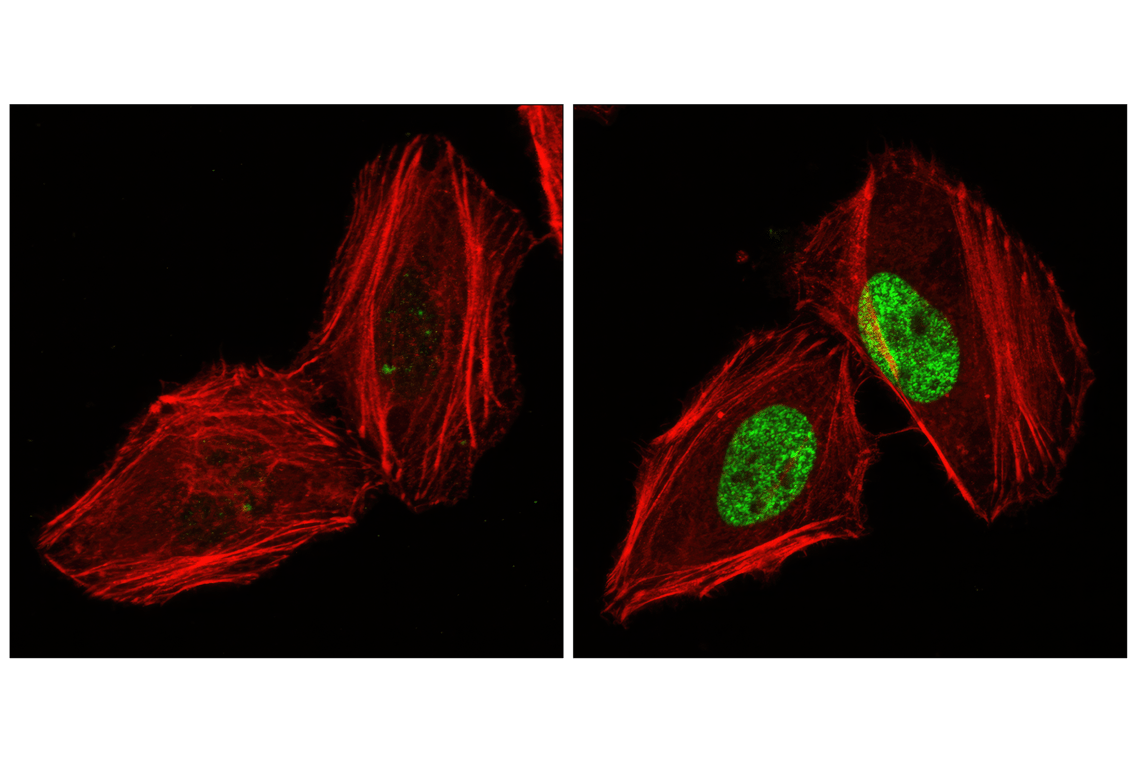
参考图片
Flow cytometric analysis of HeLa cells, untreated (blue) or treated with UV (100 mJ, 2hr recovery; green) using Phospho-H2A.X (Ser139) (20E3) Rabbit mAb (solid lines) or concentration-matched Rabbit (DA1E) mAb IgG XP® isotype control #3900 (dashed lines). Anti-rabbit IgG (H+L), F(ab')2 Fragment (Alexa Fluor® 488 Conjugate) #4412 was used as a secondary antibody.
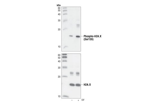
Western blot analysis of extracts from untreated or UV-treated 293 cells, using Phospho-Histone H2A.X (Ser139) (20E3) Rabbit mAb (upper) or Histone H2A.X Antibody #2595 (lower).
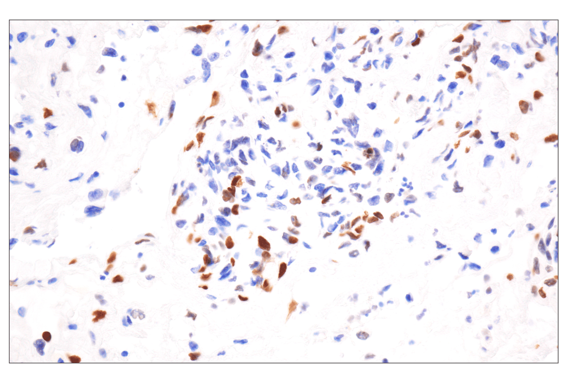
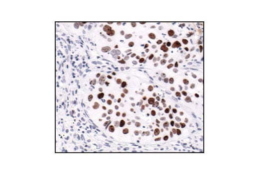
Immunohistochemical analysis of paraffin-embedded human ovarian clear cell carcinoma using Phospho-Histone H2A.X (Ser139) (20E3) Rabbit mAb performed on the Leica BOND Rx.
Immunohistochemical analysis of paraffin-embedded human prostate adenocarcinoma using Phospho-Histone H2A.X (Ser139) (20E3) Rabbit mAb performed on the Leica BOND Rx.
Immunohistochemical analysis of paraffin-embedded human lung carcinoma, using Phospho-Histone H2A.X (Ser139) (20E3) Rabbit mAb, showing nuclear localization.
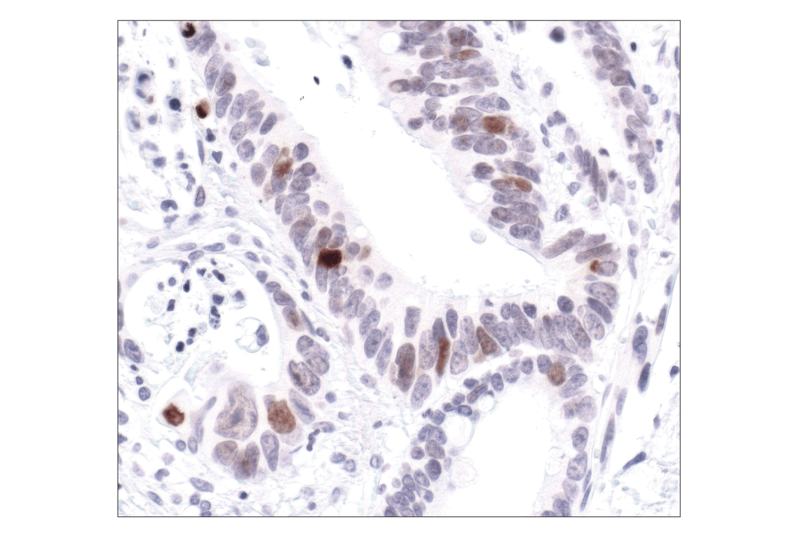
Immunohistochemical analysis of paraffin-embedded human colon carcinoma, using Phospho-Histone H2A.X (Ser139) (20E3) Rabbit mAb.
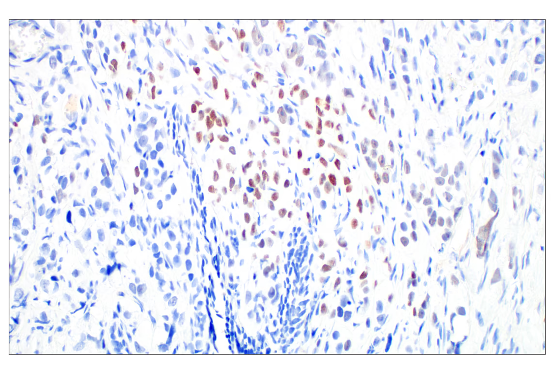
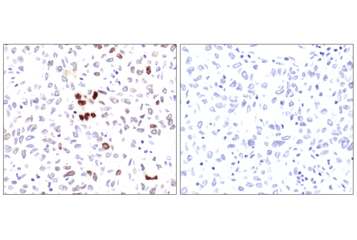
Immunohistochemical analysis of paraffin-embedded human lung carcinoma untreated (left) or lambda-phosphatase-treated (right), using Phospho-Histone H2A.X (Ser139) (20E3) Rabbit mAb.
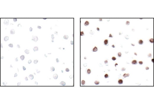
Immunohistochemical analysis of paraffin-embedded HT-29 cells untreated (left) or UV-treated (right), using Phospho-Histone H2A.X (Ser139) (20E3) Rabbit mAb.
Confocal immunofluorescent analysis of HeLa cells, untreated (left) or UV-treated (right), using Phospho-Histone H2A.X (Ser139) (20E3) Rabbit mAb (green). Actin filaments have been labeled with DY-554 phalloidin (red).