


 全部商品分类
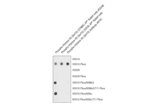


 用小程序,查商品更便捷


Monoclonal antibody is produced by immunizing animals with a synthetic phosphopeptide corresponding to residues surrounding Ser10 of human histone H3.
Product Usage Information
| Application | Dilution |
|---|---|
| Western Blotting | 1:1000 |
| Immunofluorescence (Immunocytochemistry) | 1:800 |
| Flow Cytometry (Fixed/Permeabilized) | 1:1600 |
Specificity/Sensitivity
Species Reactivity:
Human, Mouse, Rat, Monkey, Zebrafish
Supplied in 10 mM sodium HEPES (pH 7.5), 150 mM NaCl, 100 µg/ml BSA, 50% glycerol and less than 0.02% sodium azide. Store at –20°C. Do not aliquot the antibody.
For a carrier free (BSA and azide free) version of this product see product #81321.
参考图片
Peptide dot blot analysis demonstrating Phospho-Histone H3 (Ser10) (D2C8) XP® Rabbit mAb antibody specificity. Antibody binding to pre-coated histone H3 peptides is shown using Phospho-Histone H3 (Ser10) (D7N8E) XP® Rabbit mAb #53348,Phospho-Histone H3 (Ser10) (D2C8) XP® Rabbit mAb #3377, and Phospho-Histone H3 (Ser10) Antibody #9701. As shown, Phospho-Histone H3 (Ser10) (D2C8) XP® Rabbit mAb detects histone H3 phosphorylated on Ser10; however, this antibody does not detect histone H3 phosphorylated on Ser10 when Lys9 is acetylated or methylated. In addition, this antibody does not cross-react with histone H3 phosphorylated on Ser28.
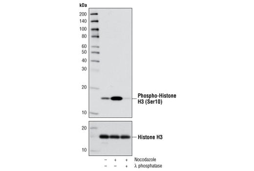
Western blot analysis of extracts from HeLa cells, either untreated or treated with nocodazole (100 ng/ml for 18 hours), using Phospho-Histone H3 (Ser10) (D2C8) XP® Rabbit mAb #3377 (upper) or Histone H3 Antibody #9715 (lower). Phospho-specificity of the antibody is shown by further treatment of the lysate with λ phosphatase.
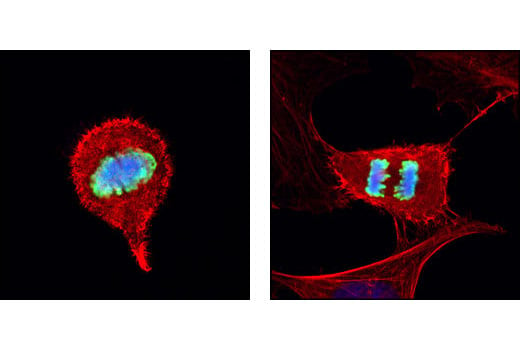
Confocal immunofluorescent analysis of HeLa cells using Phospho-Histone H3 (Ser10) (D2C8) XP® Rabbit mAb (green). Actin filaments have been labeled with DY-554 phalloidin (red). Blue pseudocolor = DRAQ5® #4084 (fluorescent DNA dye).
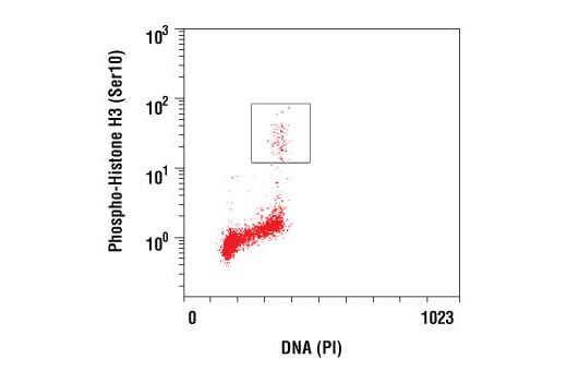
Flow cytometric analysis of Jurkat cells using Phospho-Histone H3 (Ser10) (D2C8) XP® Rabbit mAb versus propidium iodide (DNA content). The boxed population indicates Phospho-Histone H3 (Ser10) positive cells.