


 全部商品分类
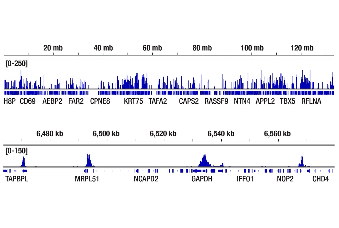


Tri-Methyl-Histone H3 (Lys4) (C42D8) Rabbit mAb

 下载产品说明书 下载COA 下载SDS
 用小程序,查商品更便捷


 收藏
收藏
 对比
对比 咨询
咨询
Monoclonal antibody is produced by immunizing animals with a synthetic peptide corresponding to the amino terminus of histone H3 in which Lys4 is tri-methylated.
Product Usage Information
For optimal ChIP and ChIP-seq results, use 10 μl of antibody and 10 μg of chromatin (approximately 4 x 10⁶ cells) per IP. This antibody has been validated using SimpleChIP® Enzymatic Chromatin IP Kits. The CUT&RUN dilution was determined using CUT&RUN Assay Kit #86652. The CUT&Tag dilution was determined using CUT&Tag Assay Kit #77552.
| Application | Dilution |
|---|---|
| Western Blotting | 1:1000 |
| Immunohistochemistry (Paraffin) | 1:1000 - 1:4000 |
| Immunofluorescence (Immunocytochemistry) | 1:200 - 1:800 |
| Flow Cytometry (Fixed/Permeabilized) | 1:400 - 1:1600 |
| Chromatin IP | 1:50 |
| Chromatin IP-seq | 1:50 |
| CUT&RUN | 1:50 |
| CUT&Tag | 1:50 |
Specificity/Sensitivity
Species Reactivity:
Human, Mouse, Rat, Monkey, D. melanogaster, S. cerevisiae
Supplied in 10 mM sodium HEPES (pH 7.5), 150 mM NaCl, 100 µg/ml BSA, 50% glycerol and less than 0.02% sodium azide. Store at –20°C. Do not aliquot the antibody.
For a carrier free (BSA and azide free) version of this product see product #19776.
参考图片
CUT&Tag was performed with HCT 116 cells and Tri-Methyl-Histone H3 (Lys4) (C42D8) Rabbit mAb, using CUT&Tag Assay Kit #77552. DNA library was prepared using CUT&Tag Dual Index Primers and PCR Master Mix for Illumina Systems #47415. The figures show binding across chromosome 12 (upper), including GAPDH (lower), a known target gene of H3K4me3 (see our ChIP-qPCR figure).
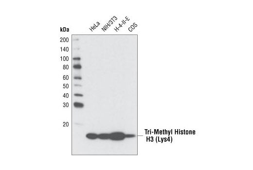
Western blot analysis of various cell types using Tri-Methyl Histone H3 (Lys4) (C42D8) Rabbit mAb.
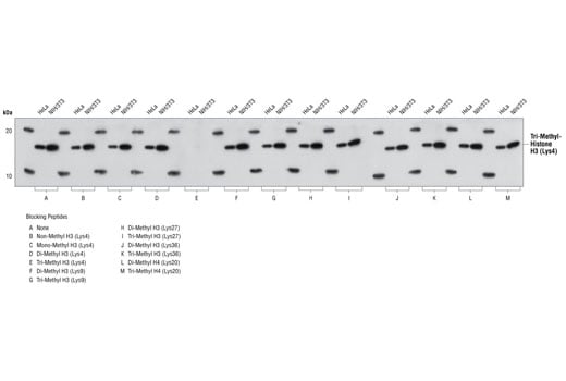
Antibody specificity was determined by Western blotting. HeLa and NIH/3T3 cell lysates were probed with Tri-Methyl Histone H3 (Lys4) (C42D8) Rabbit mAb (Panel A) or Tri-Methyl Histone H3 (Lys4) Rabbit mAb pre-adsorbed with 1.5 μM of various competitor peptides (Panels B-M). As shown, only the tri-methyl histone H3 (Lys4) peptide competed away binding of the antibody.
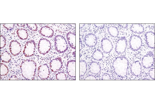
Immunohistochemical analysis of paraffin-embedded human colon using Tri-Methyl-Histone H3 (K4) (C42D8) Rabbit mAb in the presence of non-methyl peptide (left) or K4 tri-methyl peptide (right).
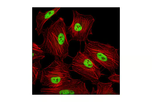
Confocal immunofluorescent analysis of HeLa cells using Tri-Methyl-Histone H3 (Lys4) (C42D8) Rabbit mAb (green). Actin filaments have been labeled with Alexa Fluor® 555 phalloidin (red).
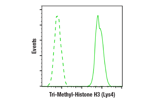
Flow cytometric analysis of HeLa cells using Tri-Methyl-Histone H3 (Lys4) (C42D8) Rabbit mAb (solid line) compared to concentration-matched Rabbit (DA1E) mAb IgG XP® Isotype Control #3900 (dashed line). Anti-rabbit IgG (H+L), F(ab')2 Fragment (Alexa Fluor® 488 Conjugate) #4412 was used as a secondary antibody.
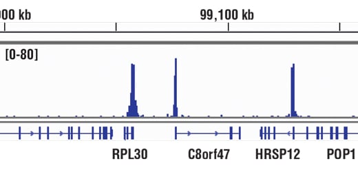
Chromatin immunoprecipitations were performed with cross-linked chromatin from HCT116 cells and Tri-Methyl-Histone H3 (Lys4) (C42D8) Rabbit mAb, using SimpleChIP® Enzymatic Chromatin IP Kit (Magnetic Beads) #9003. DNA Libraries were prepared using DNA Library Prep Kit for Illumina® (ChIP-seq, CUT&RUN) #56795. The figure shows binding across RPL30, a known target gene of H3K4me3 (see additional figure containing ChIP-qPCR data).
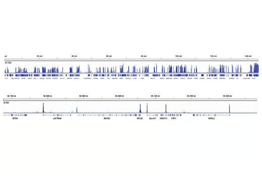
Chromatin immunoprecipitations were performed with cross-linked chromatin from HCT116 cells and Tri-Methyl-Histone H3 (Lys4) (C42D8) Rabbit mAb, using SimpleChIP® Enzymatic Chromatin IP Kit (Magnetic Beads) #9003. DNA Libraries were prepared using DNA Library Prep Kit for Illumina® (ChIP-seq, CUT&RUN) #56795. The figure shows binding across chromosome 8 (upper), including RPL30 (lower), a known target gene of H3K4me3 (see additional figure containing ChIP-qPCR data).
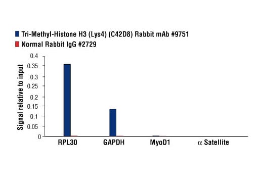
Chromatin immunoprecipitations were performed with cross-linked chromatin from HeLa cells and either Tri-Methyl-Histone H3 (Lys4) (C42D8) Rabbit mAb or Normal Rabbit IgG #2729, using SimpleChIP® Enzymatic Chromatin IP Kit (Magnetic Beads) #9003. The enriched DNA was quantified by real-time PCR using SimpleChIP® Human RPL30 Exon 3 Primers #7014, SimpleChIP® Human GAPDH Exon 1 Primers #5516, SimpleChIP® Human MyoD1 Exon 1 Primers #4490, and SimpleChIP® Human α Satellite Repeat Primers #4486. The amount of immunoprecipitated DNA in each sample is represented as signal relative to the total amount of input chromatin, which is equivalent to one.
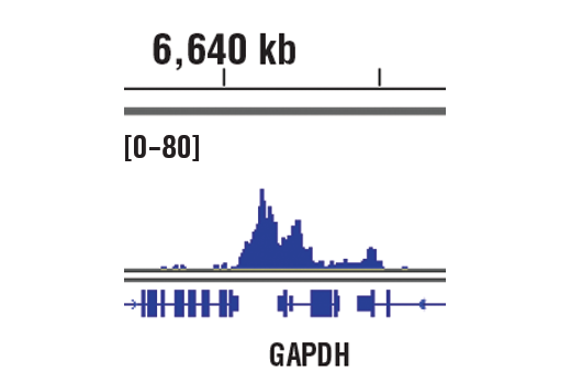
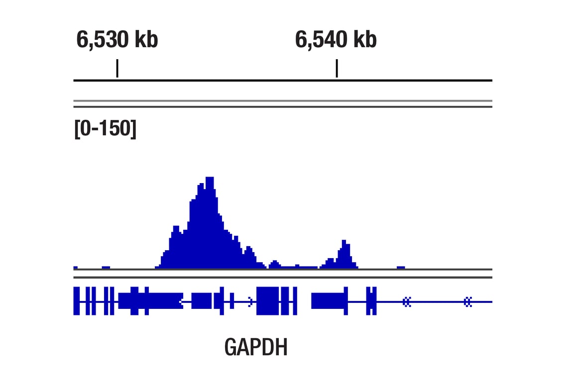
CUT&RUN was performed with HCT 116 cells and Tri-Methyl-Histone H3 (Lys4) (C42D8) Rabbit mAb, using CUT&RUN Assay Kit #86652. DNA Libraries were prepared using DNA Library Prep Kit for Illumina® (ChIP-seq, CUT&RUN) #56795. The figures show binding across GAPDH, a known target gene of H3K4me3 (see additional figure containing CUT&RUN-qPCR data).
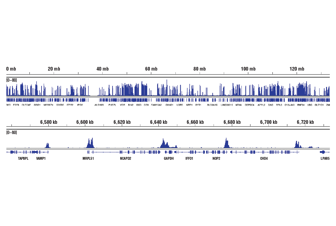
CUT&RUN was performed with HCT 116 cells and Tri-Methyl-Histone H3 (Lys4) (C42D8) Rabbit mAb, using CUT&RUN Assay Kit #86652. DNA Libraries were prepared using DNA Library Prep Kit for Illumina® (ChIP-seq, CUT&RUN) #56795. The figures show binding across chromosome 12 (upper), including GAPDH (lower), a known target gene of H3K4me3 (see additional figure containing CUT&RUN-qPCR data).
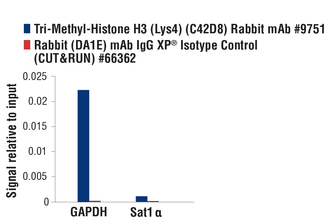
CUT&RUN was performed with HCT 116 cells and Tri-Methyl-Histone H3 (Lys4) (C42D8) Rabbit mAb or Rabbit (DA1E) mAb IgG XP® Isotype Control (CUT & RUN) #66362, using CUT&RUN Assay Kit #86652. The enriched DNA was quantified by real-time PCR using SimpleChIP® Human GAPDH Exon 1 Primers #5516 and SimpleChIP® Human α Satellite Repeat Primers #4486. The amount of immunoprecipitated DNA in each sample is represented as signal relative to the total amount of input chromatin, which is equivalent to one.
CUT&Tag was performed with HCT 116 cells and Tri-Methyl-Histone H3 (Lys4) (C42D8) Rabbit mAb, using CUT&Tag Assay Kit #77552. DNA library was prepared using CUT&Tag Dual Index Primers and PCR Master Mix for Illumina Systems #47415. The figure shows binding across GAPDH, a known target gene of H3K4me3 (see our ChIP-qPCR figure).