


 下载产品说明书 下载SDS
 用小程序,查商品更便捷


 收藏
收藏
 对比
对比 咨询
咨询
Product Usage Information
For optimal ChIP and ChIP-seq results, use 10 μl of antibody and 10 μg of chromatin (approximately 4 x 10⁶ cells) per IP. This antibody has been validated using SimpleChIP® Enzymatic Chromatin IP Kits. The CUT&RUN dilution was determined using CUT&RUN Assay Kit #86652. The CUT&Tag dilution was determined using 100,000 cells in a total reaction volume of 100 μl.
| Application | Dilution |
|---|---|
| Western Blotting | 1:1000 |
| Immunohistochemistry (Paraffin) | 1:1000 - 1:4000 |
| Immunofluorescence (Immunocytochemistry) | 1:200 - 1:800 |
| Flow Cytometry (Fixed/Permeabilized) | 1:400 - 1:1600 |
| Chromatin IP | 1:50 |
| Chromatin IP-seq | 1:50 |
| CUT&RUN | 1:50 |
| CUT&Tag | 1:50 |
Specificity/Sensitivity
物种反应性:
人, 小鼠, 大鼠, 猴, 黑腹果蝇 , 酿酒酵母
参考图片
Confocal immunofluorescent analysis of HeLa cells using Tri-Methyl-Histone H3 (Lys4) (C42D8) Rabbit mAb (green). Actin filaments have been labeled with Alexa Fluor® 555 phalloidin (red).
使用Tri-Methyl-Histone H3 (Lys4) (C42D8) Rabbit mAb (绿色),共聚焦免疫荧光分析HeLa细胞。Alexa Fluor® 555 phalloidin标记微丝蛋白(红色)。
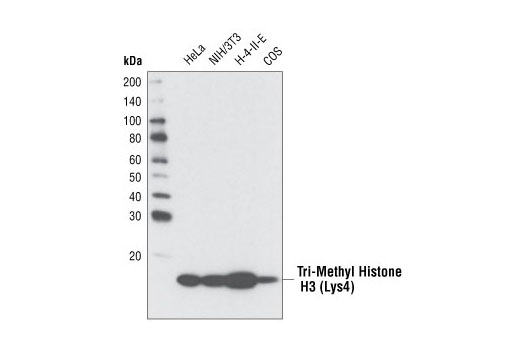
Western blot analysis of various cell types using Tri-Methyl Histone H3 (Lys4) (C42D8) Rabbit mAb.
使用Tri-Methyl Histone H3 (Lys4) (C42D8) Rabbit mAb,免疫印迹(Western blot)分析不同细胞中Tri-Methyl-Histone H3 (Lys4)的蛋白水平。
Antibody specificity was determined by Western blotting. HeLa and NIH/3T3 cell lysates were probed with Tri-Methyl Histone H3 (Lys4) (C42D8) Rabbit mAb (Panel A) or Tri-Methyl Histone H3 (Lys4) Rabbit mAb pre-adsorbed with 1.5 μM of various competitor peptides (Panels B-M). As shown, only the tri-methyl histone H3 (Lys4) peptide competed away binding of the antibody.
抗体特异性通过免疫印迹(Western blot)确定。HeLa和NIH/3T3细胞提取物的探针使用只有Tri-Methyl Histone H3 (Lys4) (C42D8) Rabbit mAb (图A)或Tri-Methyl Histone H3 (Lys4) Rabbit mAb,并且其提前孵育了1.5 μM various competitor peptides (图B-I)。如图所示,仅tri-methyl histone H3 (Lys4) peptide竞争离开该抗体的结合。
Chromatin immunoprecipitations were performed with cross-linked chromatin from 4 x 10⁶ HeLa cells and either 10 μl of Tri-Methyl-Histone H3 (Lys4) (C42D8) Rabbit mAb or 2 μl of Normal Rabbit IgG #2729, using SimpleChIP® Enzymatic Chromatin IP Kit (Magnetic Beads) #9003. The enriched DNA was quantified by real-time PCR using SimpleChIP® Human RPL30 Exon 3 Primers #7014, SimpleChIP® Human GAPDH Exon 1 Primers #5516, SimpleChIP® Human MyoD1 Exon 1 Primers #4490, and SimpleChIP® Human α Satellite Repeat Primers #4486. The amount of immunoprecipitated DNA in each sample is represented as signal relative to the total amount of input chromatin, which is equivalent to one.
使用SimpleChIP®Enzymatic Chromatin IP Kit (Magnetic Beads) #9003,用4 x 10⁶ HeLa细胞的交联染色质以及10 µl Tri-Methyl-Histone H3 (Lys4) (C42D8) Rabbit mAb或2 µl Normal Rabbit IgG #2729进行染色质免疫沉淀实验。使用SimpleChIP® Human RPL30 Exon 3 Primers #7014、SimpleChIP® Human GAPDH Exon 1 Primers #5516、SimpleChIP® Human MyoD1 Exon 1 Primers #4490和SimpleChIP® Human α Satellite Repeat Primers #4486,浓缩的DNA通过real-time PCR定量。在每个样品中免疫沉淀DNA的数量被当做一个相对于总input chromatin的数量的信号,相当于一。
Immunohistochemical analysis of paraffin-embedded human colon using Tri-Methyl-Histone H3 (K4) (C42D8) Rabbit mAb in the presence of non-methyl peptide (left) or K4 tri-methyl peptide (right).
Flow cytometric analysis of human whole blood cells using Tri-Methyl-Histone H3 (Lys4) (C42D8) Rabbit mAb (blue) and Rabbit (DA1E) mAb IgG XP® Isotype Control #3900 (red). Anti-rabbit IgG (H+L), F(ab')2 Fragment (Alexa Fluor® 647 Conjugate) #4414 was used as a secondary antibody. Analysis was performed on cells in the lymphocyte gate.

 危险品化学品经营许可证(不带存储) 许可证编号:沪(杨)应急管危经许[2022]202944(QY)
  营业执照(三证合一)