


 全部商品分类
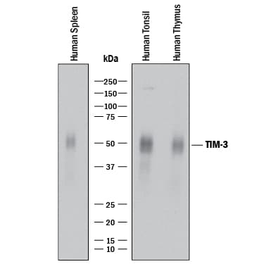


 用小程序,查商品更便捷


Scientific Data
 View Larger
Detection of Human TIM‑3 by Western Blot. Western blot shows lysates of human spleen tissue, human tonsil tissue, and human thymus tissue. PVDF membrane was probed with 2 µg/mL of Rabbit Anti-Human TIM-3 Monoclonal Antibody (Catalog # MAB23652) followed by HRP-conjugated Anti-Rabbit IgG Secondary Antibody (Catalog # HAF008). A specific band was detected for TIM-3 at approximately 45-70 kDa (as indicated). This experiment was conducted under reducing conditions and using Immunoblot Buffer Group 1.
 View Larger
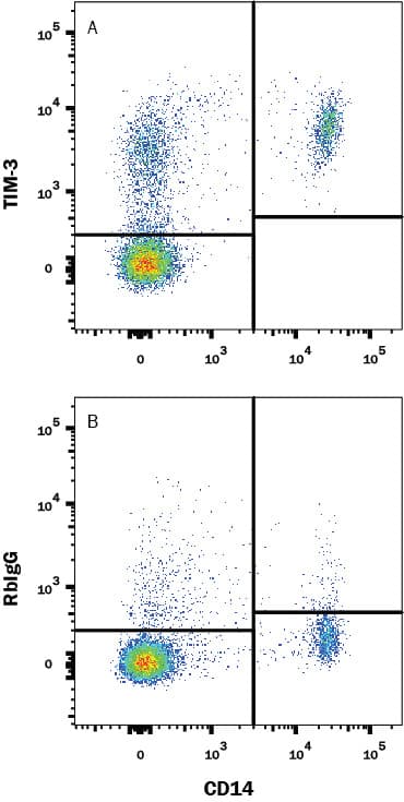
Detection of Tim-3 in Human PBMC by Flow Cytometry. Human PBMC were stained with (A) Rabbit Anti-Human Tim-3 Monoclonal Antibody (Catalog # MAB23652) or (B) Normal Rabbit IgG Control Antibody (Catalog # MAB1050) followed by PE-conjugated Goat anti-Rabbit IgG Secondary Antibody (Catalog # F0110) and Mouse anti-Human CD14 APC-conjugated Monoclonal Antibody (Catalog # FAB3832A). View our protocol for Staining Membrane-associated Proteins.
.") View Larger
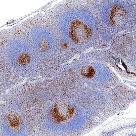
View LargerTIM‑3 in Human Tonsil. TIM-3 was detected in immersion fixed paraffin-embedded sections of human tonsil using Rabbit Anti-Human TIM-3 Monoclonal Antibody (Catalog # MAB23652) at 3 µg/mL for 1 hour at room temperature followed by incubation with the Anti-Rabbit IgG VisUCyte™ HRP Polymer Antibody (Catalog # VC003). Before incubation with the primary antibody, tissue was subjected to heat-induced epitope retrieval using Antigen Retrieval Reagent-Basic (Catalog # CTS013). Tissue was stained using DAB (brown) and counterstained with hematoxylin (blue). Specific staining was localized to cytoplasm in lymphocytes. View our protocol for IHC Staining with VisUCyte HRP Polymer Detection Reagents.
Human TIM-3 Antibody Summary
Met1-Arg200
Accession # Q8TDQ0
Applications
Please Note: Optimal dilutions should be determined by each laboratory for each application. General Protocols are available in the Technical Information section on our website.
Background: TIM-3
TIM-3 (T cell immunoglobulin and mucin domain-3) is a 60 kDa member of the TIM family of immune regulating molecules. TIMs are type I transmembrane glycoproteins with one Ig-like V-type domain and a Ser/Thr-rich mucin stalk (1-3). There are three TIM genes in human and eight in mouse. Mature human TIM-3 consists of a 181 amino acid (aa) extracellular domain (ECD), a 21 aa transmembrane segment, and a 78 aa cytoplasmic tail (4). An alternately spliced isoform is truncated following a short substitution after the Ig-like domain. Within the ECD, human TIM-3 shares 58% aa sequence identity with mouse and rat TIM-3. TIM-3 is expressed on the surface of effector T cells (CD4+ Th1 and CD8+ Tc1) but not on helper T cells (CD4+ Th2 and CD8+ Tc2) (4, 5). NK cells appear to transcribe the highest amounts of Tim-3 among lymphocytes, and when Tim-3 was cross-linked with antibodies it suppressed NK cell-mediated cytotoxicity (6). In chronic inflammation, autoimmune disorders, and some cancers, TIM-3 is upregulated on several other hematopoietic cell types. It also occurs on hippocampal neurons (7-10). The Ig domain of TIM-3 interacts with a ligand on resting but not activated Th1 and Th2 cells (5, 11). The glycosylated Ig domain of TIM-3 binds cell-associated galectin-9. This induces TIM-3 Tyr phosphorylation and pro-apoptotic signaling (8, 12). TIM-3 functions as a negative regulator of Th1 cell activity. Its blockade results in increased IFN-gamma production, Th1 cell proliferation and cytotoxicity (5, 10, 11), and regulatory T cell development (5). TIM-3 inhibits the antitumor efficacy of DNA vaccines and chemotherapy by binding to the damage-associated molecular pattern molecule, HMGB1 (13).
- Anderson, A.C. and D.E. Anderson (2006) Curr. Opin. Immunol. 18:665.
- Mariat, C. et al. (2005) Phil. Trans. R. Soc. B. 360:1681.
- Meyers, J.H. et al. (2005) Trends Mol. Med. 11:362.
- Monney, L. et al. (2002) Nature 415:536.
- Sanchez-Fueyo, A. et al. (2003) Nat. Immunol. 4:1093.
- Ndhlovu, L. et al. (2012) Blood 119:3734.
- Wiener, Z. et al. (2007) J. Invest. Dermatol. 127:906.
- van de Weyer, P.S. et al. (2006) Biochem. Biophys. Res. Commun. 351:571.
- Gielen, A.W. et al. (2005) J. Neuroimmunol. 164:93.
- Oikawa, T. et al. (2006) J. Immunol. 177:4281.
- Sabatos, C.A. et al. (2003) Nat. Immunol. 4:1102.
- Zhu, C. et al. (2005) Nat. Immunol. 6:1245.
- Chiba, S. et al. (2012) Nat. Immunol. 13:832.
Preparation and Storage
- 12 months from date of receipt, -20 to -70 °C as supplied.
- 1 month, 2 to 8 °C under sterile conditions after reconstitution.
- 6 months, -20 to -70 °C under sterile conditions after reconstitution.