


 全部商品分类
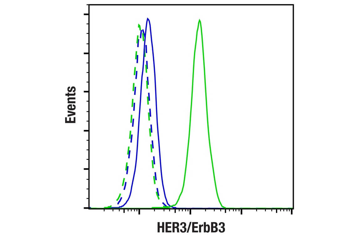


HER3/ErbB3 (D22C5) XP ® Rabbit mAb

 下载产品说明书 下载COA 下载SDS
 用小程序,查商品更便捷


 收藏
收藏
 对比
对比 咨询
咨询
Monoclonal antibody is produced by immunizing animals with recombinant protein corresponding to the carboxy terminus of human ErbB3 protein.
Product Usage Information
| Application | Dilution |
|---|---|
| Western Blotting | 1:1000 |
| Simple Western™ | 1:50 - 1:250 |
| Immunoprecipitation | 1:50 |
| IHC Leica Bond | 1:50 |
| Immunohistochemistry (Paraffin) | 1:125 - 1:500 |
| Immunofluorescence (Immunocytochemistry) | 1:100 - 1:200 |
| Flow Cytometry (Fixed/Permeabilized) | 1:100 - 1:400 |
Specificity/Sensitivity
Species Reactivity:
Human, Mouse
Supplied in 10 mM sodium HEPES (pH 7.5), 150 mM NaCl, 100 µg/ml BSA, 50% glycerol and less than 0.02% sodium azide. Store at –20°C. Do not aliquot the antibody.
For a carrier free (BSA and azide free) version of this product see product #60638.
参考图片
Flow cytometric analysis of MDA-MB-231 cells (blue, negative) and T-47D cells (green, positive) using HER3/ErbB3 (D22C5) XP® Rabbit mAb (solid lines) or concentration-matched Rabbit (DA1E) mAb IgG XP® Isotype Control #3900 (dashed lines). Anti-rabbit IgG (H+L), F(ab')2 Fragment (Alexa Fluor® 647 Conjugate) #4414 was used as a secondary antibody.
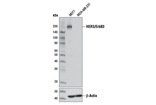
Western blot analysis of extracts from MCF7 (HER3+) and MDA-MB-231 (HER3-) cells using HER3/ErbB3 (D22C5) XP® Rabbit mAb (upper) or β-Actin (D6A8) Rabbit mAb #8457 (lower).
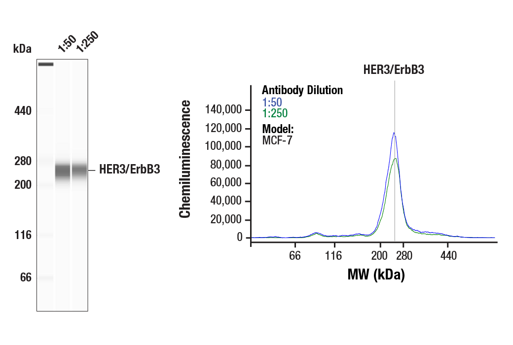
Simple Western™ analysis of lysates (1.0 mg/mL) from MCF-7 cells using HER3/ErbB3 (D22C5) XP® Rabbit mAb #12708. The virtual lane view (left) shows the target band (as indicated) at 1:50 and 1:250 dilutions of primary antibody. The corresponding electropherogram view (right) plots chemiluminescence by molecular weight along the capillary at 1:50 (blue line) and 1:250 (green line) dilutions of primary antibody. This experiment was performed under reducing conditions on the Jess™ Simple Western instrument from ProteinSimple, a BioTechne brand, using the 44-660 kDa separation module.
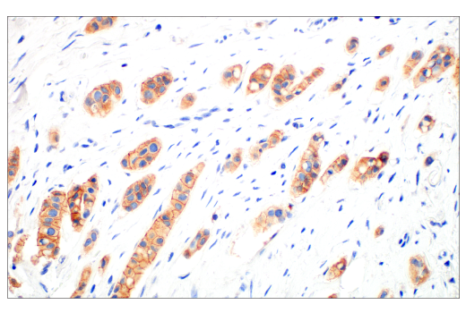
Immunohistochemical analysis of paraffin-embedded human ductal breast carcinoma using HER3/ErbB3 (D22C5) XP® Rabbit mAb performed on the Leica BOND Rx.
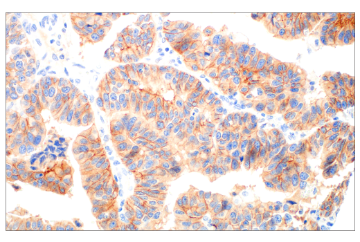
Immunohistochemical analysis of paraffin-embedded human ovarian clear cell carcinoma using HER3/ErbB3 (D22C5) XP® Rabbit mAb performed on the Leica BOND Rx.
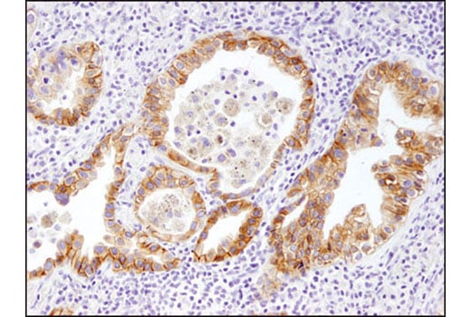
Immunohistochemical analysis of paraffin-embedded human gastric carcinoma using HER3/ErbB3 (D22C5) XP® Rabbit mAb performed on the Leica BOND Rx.
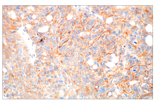
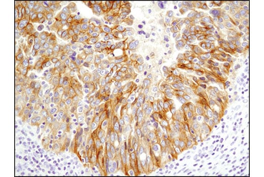
Immunohistochemical analysis of paraffin-embedded non-small cell lung carcinoma using HER3/ErbB3 (D22C5) XP® Rabbit mAb.
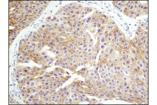
Immunohistochemical analysis of paraffin-embedded breast carcinoma using HER3/ErbB3 (D22C5) XP® Rabbit mAb.
Immunohistochemical analysis of paraffin-embedded ovarian serous adenocarcinoma using HER3/ErbB3 (D22C5) XP® Rabbit mAb.
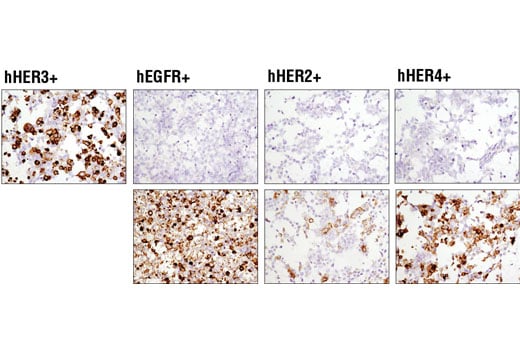
Immunohistochemical analysis of paraffin-embedded 293T cell pellets transfected with human erbB family members HER3, EGFR, HER2 and HER4 (from left to right as indicated) using HER3/ErbB3 (D22C5) XP® Rabbit mAb (top panels). Transfections were confirmed using EGF Receptor (D38B1) XP® Rabbit mAb #4267 (lower left), HER2/ErbB2 (D8F12) XP® Rabbit mAb #4290 (lower middle) and HER4/ErbB4 (111B2) Rabbit mAb #4795 (lower right).
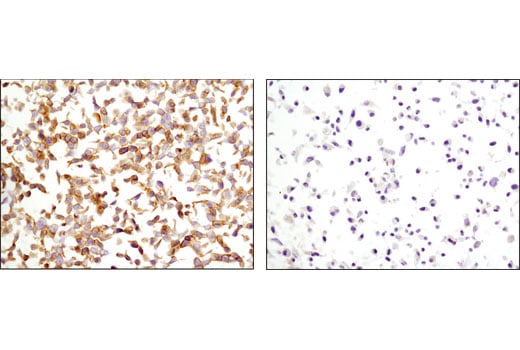
Immunohistochemical analysis of paraffin-embedded MCF7 (left) or MDA-MB-231 (right) cell pellets using HER3/ErbB3 (D22C5) XP® Rabbit mAb.
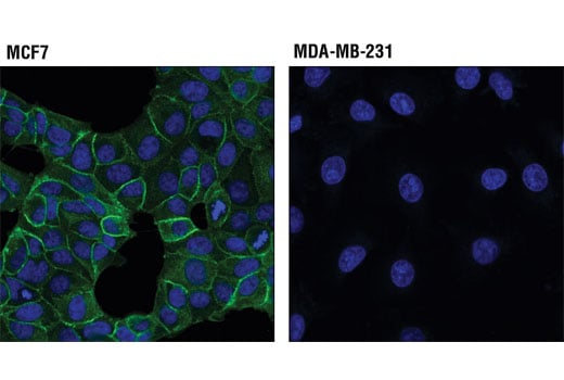
Confocal immunofluorescent analysis of MCF7 cells (HER3+, left) and MDA-MB-231 cells (HER-, right) using HER3/ErbB3 (D22C5) XP® Rabbit mAb (green). Blue pseudocolor= DRAQ5® #4084 (fluorescent DNA dye).