


 全部商品分类


Hippo Signaling Antibody Sampler Kit

 下载产品说明书 下载SDS
 用小程序,查商品更便捷


 收藏
收藏
 对比
对比 咨询
咨询
The Hippo Signaling Antibody Sampler Kit provides an economical means of detecting target proteins of the Hippo signaling pathway. The kit contains enough primary antibody to perform two western blots per primary.
参考图片
Immunohistochemical analysis of paraffin-embedded human non-small cell lung carcinoma using Phospho-YAP (Ser127) (D9W2I) Rabbit mAb in the presence of control peptide (left) or antigen-specific peptide (right).
Western blot analysis of extracts from HeLa cells (lane 1) or YAP knock-out cells (lane 2) using Phospho-YAP (Ser127) (D9W2I) Rabbit mAb #13008 (upper), and GAPDH (D6H11) XP® Rabbit mAb #5174 (lower). The absence of signal in the YAP knock-out HeLa cells confirms specificity of the antibody for YAP.
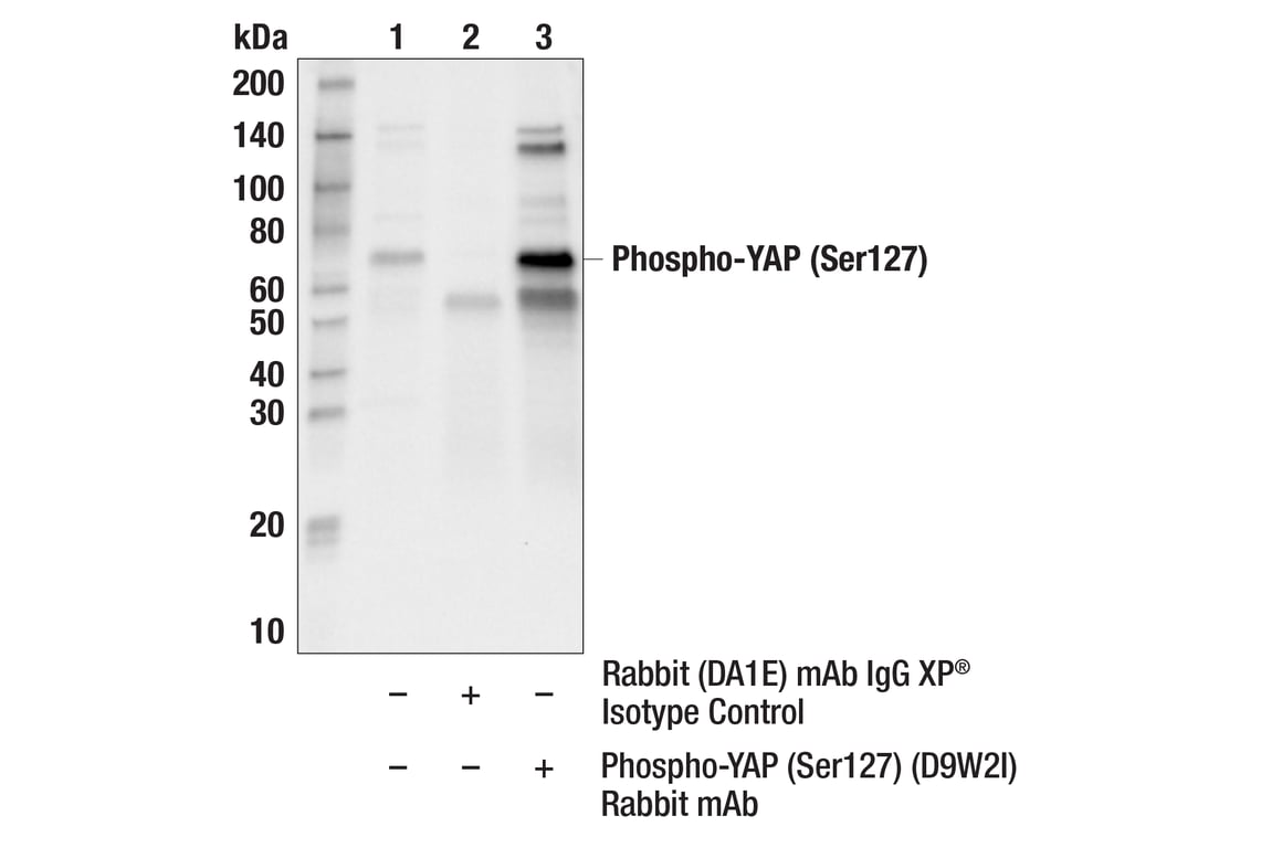
Immunoprecipitation of Phospho-YAP (Ser127) protein from C6 serum starved + Forskolin #3828 (20µM, 1 hr) cell extracts. Lane 1 is 10% input, lane 2 is Rabbit (DA1E) mAb IgG XP® Isotype Control #3900, and lane 3 is Phospho-YAP (Ser127) (D9W2I) Rabbit mAb. Western blot analysis was performed using Phospho-YAP (Ser127) (D9W2I) Rabbit mAb. Mouse Anti-rabbit IgG (Conformation Specific) (L27A9) mAb #3678 was used as a secondary antibody.
Western blot analysis of extracts from various cell lines using SAV1 (D6M6X) Rabbit mAb (upper) and β-Actin (D6A8) Rabbit mAb #8457 (lower).
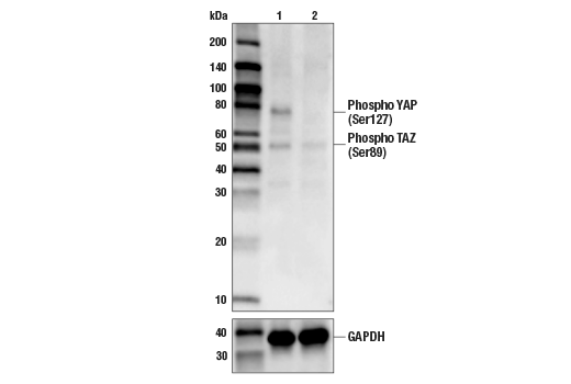
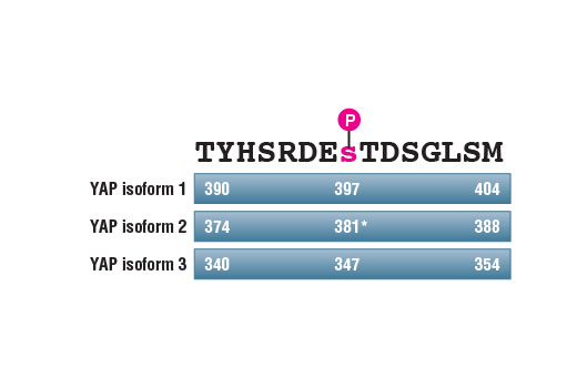
Western blot analysis of extracts from Hep G2 cells, untreated (-) or λ-phosphatase-treated (+), using Phospho-YAP (Ser397) (D1E7Y) Rabbit mAb (upper), YAP Antibody #4912 (middle), and β-Actin (D6A8) Rabbit mAb #8457 (lower). YAP protein isoform 1 Ser397 corresponds to Ser381 of YAP isoform 2, as reported by Zhao et al. (2010) Genes Dev 24, 72-85 (9).
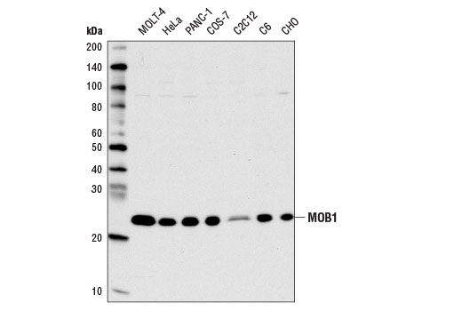
Western blot analysis of extracts from various cell lines using MOB1 (E1N9D) Rabbit mAb.
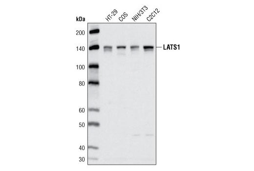
Western blot analysis of extracts from various cell lines using LATS1 (C66B5) Rabbit mAb.
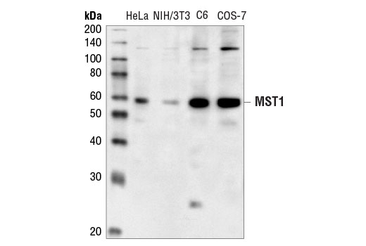
Western blot analysis of extracts from various cell lines using MST1 Antibody.
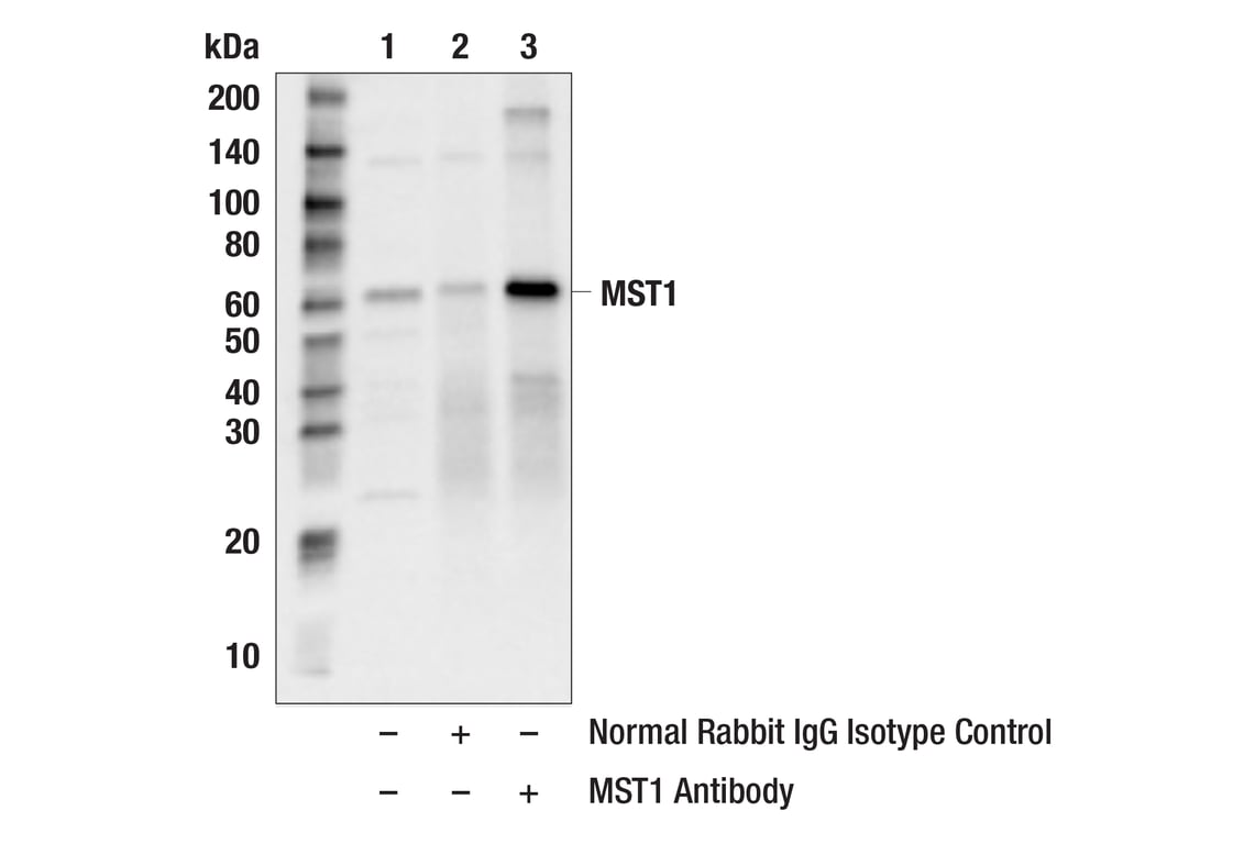
Immunoprecipitation of MST1 protein from HeLa cell extracts. Lane 1 is 10% input, lane 2 is Normal Rabbit IgG #2729, and lane 3 is MST1 Antibody. Western blot analysis was performed using MST1 Antibody. Mouse Anti-rabbit IgG (Conformation Specific) (L27A9) mAb #3678 was used as a secondary antibody.
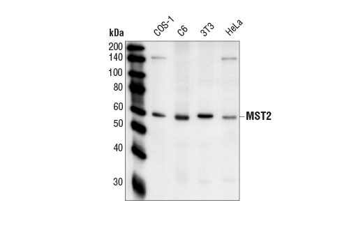
Western blot analysis of extracts from various cell lines using MST2 Antibody.
Immunoprecipitation of MST2 protein from A431 cell extracts. Lane 1 is 10% input, lane 2 is Normal Rabbit IgG #2729, and lane 3 is MST2 Antibody. Western blot analysis was performed using MST2 Antibody. Mouse Anti-rabbit IgG (Conformation Specific) (L27A9) mAb #3678 was used as a secondary antibody.
After the primary antibody is bound to the target protein, a complex with HRP-linked secondary antibody is formed. The LumiGLO® is added and emits light during enzyme catalyzed decomposition.
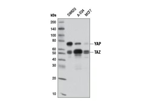
Western blot analysis of extracts from various cell lines using YAP/TAZ (D24E4) Rabbit mAb.
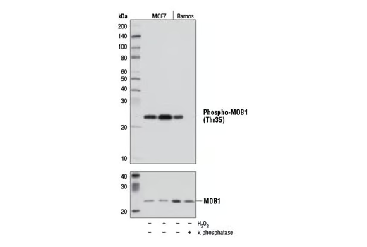
Western blot analysis of extracts from MCF7 cells, either untreated (-) or treated (+) with H2O2 (2.5 mM, 30 min) and Ramos cells, either untreated (-) or treated (+) with λ phosphatase, using Phospho-MOB1 (Thr35) (D2F10) Rabbit mAb (upper) and MOB1 Antibody #3863 (lower).
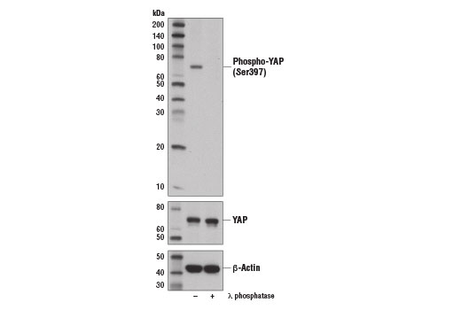
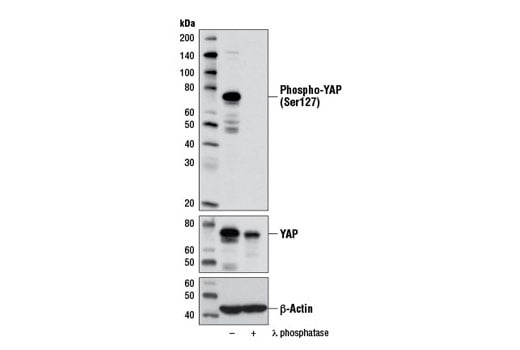
Western blot analysis of extracts from PANC-1 cells, untreated (-) or λ-phosphatase-treated (+), using Phospho-YAP (Ser127) (D9W2I) Rabbit mAb (upper), YAP Antibody #4912 (middle), and β-Actin (D6A8) Rabbit mAb #8457 (lower).
Immunoprecipitation of SAV1 protein from A-204 cell extracts using Rabbit (DA1E) mAb IgG XP® Isotype Control #3900 (lane 2) or SAV1 (D6M6X) Rabbit mAb (lane 3). Lane 1 is 10% input. Western blot analysis was performed using SAV1 (D6M6X) Rabbit mAb.
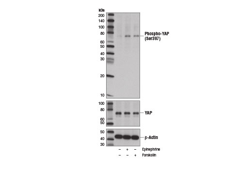
Western blot analysis of extracts from MDA-MB-231 cells, vehicle treated (-) or treated with epinephrine (10 μM, 60 min; +) or Forskolin #3828 (10 μm, 60 min; +), using Phospho-YAP (Ser397) (D1E7Y) Rabbit mAb (upper), YAP Antibody #4912 (middle), and β-Actin (D6A8) Rabbit mAb #8547 (lower). YAP protein isoform 1 Ser397 corresponds to Ser381 of YAP isoform 2, as reported by Zhao, B. et al. (2010) Genes Dev 24, 72-85 (9).
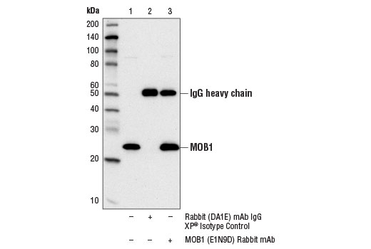
Immunoprecipitation of MOB1 protein from HeLa cell extracts using Rabbit (DA1E) mAb IgG XP® Isotype Control #3900 (lane 2) or MOB1 (E1N9D) Rabbit mAb (lane 3). Lane 1 is 10% input. Western blot analysis was performed using MOB1 (E1N9D) Rabbit mAb.
Western blot analysis of extracts from HeLa cells treated with Staurosporine #9953 for the indicated times using MST2 Antibody.
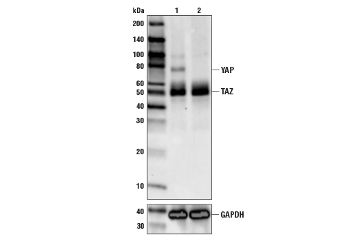
Western blot analysis of extracts from HeLa cells (lane 1) or YAP knock-out cells (lane 2) using YAP/TAZ (D24E4) Rabbit mAb #8418 (upper), and GAPDH (D6H11) XP® Rabbit mAb #5174 (lower). The absence of signal in the YAP knock-out HeLa cells confirms specificity of the antibody for YAP.
Immunohistochemical analysis of paraffin-embedded human lung carcinoma using Phospho-MOB1 (Thr35) (D2F10) Rabbit mAb.
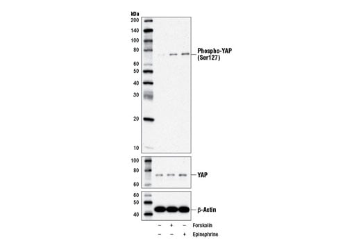
Western blot analysis of MDA-MB-231 cells, vehicle-treated (-) or treated with Forskolin #3828 (10 μM, 60 min; +) or epinephrine (10 μM, 60 min; +), using Phospho-YAP (Ser127) (D9W2I) Rabbit mAb (upper), YAP Antibody #4912 (middle), and β-Actin (D6A8) Rabbit mAb #8457 (lower). Note the induction of YAP (Ser127) phosphorylation after treatment with forskolin or epinephrine, consistent with the findings reported in Xu et al. (2012) [9].
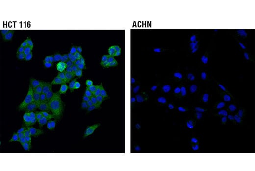
Confocal immunofluorescent analysis of HCT 116 (higher expressing, left) and ACHN (lower expressing, right) cells using SAV1 (D6M6X) Rabbit mAb (green). Blue pseudocolor = DRAQ5® #4084 (fluorescent DNA dye).
Western blot analysis of extracts from various cell lines using Phospho-YAP (Ser397) (D1E7Y) Rabbit mAb.
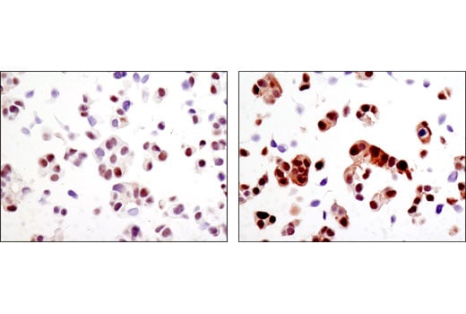
Immunohistochemical analysis of paraffin-embedded human ovarian carcinoma control (left) or lambda phosphatase-treated (right) using Phospho-MOB1 (Thr35) (D2F10) Rabbit mAb.
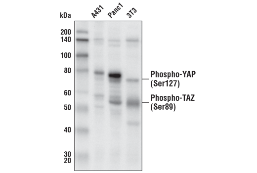
Western blot analysis of extracts from various cell lines using Phospho-YAP (Ser127) (D9W2I) Rabbit mAb.
Schematic diagram showing annotation of the amino acid sequence surrounding Ser397 in different human YAP isoforms. Asterisk (*) indicates annotation of the phosphorylation site as described in Zhao et al. 2010 [9].
Immunohistochemical analysis of paraffin-embedded MCF7 cell pellets, control (left) or H2O2-treated (right), using Phospho-MOB1 (Thr35) (D2F10) Rabbit mAb.
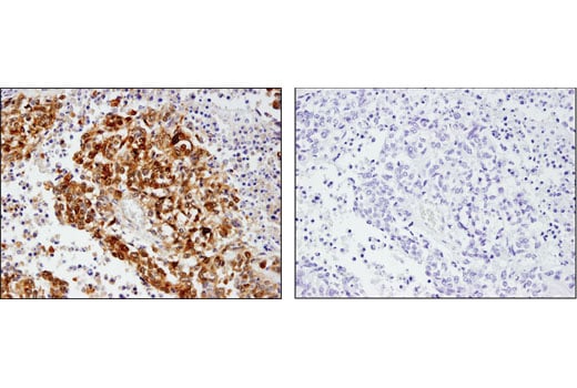
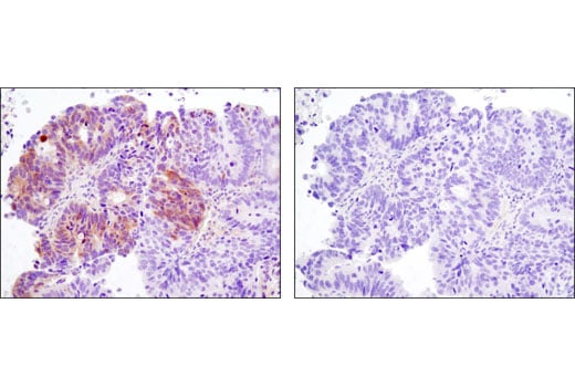
Immunohistochemical analysis of paraffin-embedded human colon adenocarcinoma, control (left) or λ-phosphatase treated (right), using Phospho-YAP (Ser127) (D9W2I) Rabbit mAb.
Immunohistochemical analysis of paraffin-embedded cell pellets, A-204 (left) and RL (right), using Phospho-YAP (Ser127) (D9W2I) Rabbit mAb.