


 全部商品分类


BD Pharmingen™ PE Mouse Anti-Human HLA-DR

 下载产品说明书 下载SDS
 用小程序,查商品更便捷


 收藏
收藏
 对比
对比 咨询
咨询

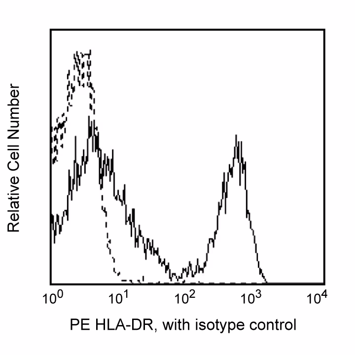
参考图片
Flow cytometric analysis of HLDA-DR expression on Rhesus macaque (Macaca mulatta) peripheral blood lymphocytes. Rhesus whole blood was stained with either PE Mouse IgG2a, κ Isotype Control (Cat. No. 556653; dashed line histogram) or PE Mouse Anti-Human HLA-DR (Cat. No. 556644/555812/560943; solid line histogram). Erythrocytes were lysed with BD FACS™ Lysing Solution (Cat. No. 349202). Fluorescent histograms were derived from forward and side light-scatter characteristics of viable lymphocytes.
Flow cytometric analysis of HLDA-DR expression on Rhesus macaque (Macaca mulatta) peripheral blood lymphocytes. Rhesus whole blood was stained with either PE Mouse IgG2a, κ Isotype Control (Cat. No. 556653; dashed line histogram) or PE Mouse Anti-Human HLA-DR (Cat. No. 556644/555812/560943; solid line histogram). Erythrocytes were lysed with BD FACS™ Lysing Solution (Cat. No. 349202). Fluorescent histograms were derived from forward and side light-scatter characteristics of viable lymphocytes.