首页
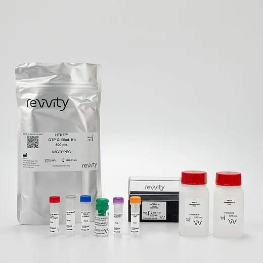
HTRF GTP GI BIND. KIT - 500 PTS


品牌: Revvity
 下载产品说明书
 下载SDS
下载SDS 用小程序,查商品更便捷


 收藏
收藏
 对比
对比 咨询
咨询
产品介绍
产品信息
Overview
The GTP Gi binding assay detects upstream Gi protein activity at the receptor, upon GPCR stimulation in membrane extracts. This HTRF-based non-isotopic assay is simple, fast, and has a no-wash formatThe assay is intended for GPCR model studies, and compound pharmacological identification and characterization.
Specifications
| Assay Points | 100 |
|---|---|
| Assay Target Type | Kit |
| Assay Technology | HTRF |
| Brand | HTRF |
| Quantity | 1 |
| Therapeutic Area | Cardiovascular Infectious Diseases Inflammation Metabolism/Diabetes NASH/Fibrosis Neuroscience Oncology & Inflammation Rare Diseases |
| Unit Size | 100 Assay Points |
Video gallery
How it works
GTP Gi binding assay principle
The GTP Gi binding assay uses a cryptate-labelled GTP analog (donor) and a d2-labelled anti-Gi antibody (acceptor). The GTP analog specifically binds into the GTP-binding pocket of active G proteins, while the d2-labelled antibody is specific to the Gαi subunit of G proteins. GPCR activation in presence of the two detection reagents brings the donor and acceptor into proximity, which triggers a FRET signal with an intensity proportional to the concentration of active Gi protein in the sample. GTP gamma S, a non-hydrolysable GTP analog, is used to displace the donor from the GTP-binding pocket and extinguish FRET to determine the assay non-specific signal an ensure its accuracy.

GTP Gi binding-plate assay protocol
The GTP Gi binding assay is performed in a miniaturized format using 20 µl final volume, and features a streamlined 4-step reagent addition protocol where stimulation/compounds, agonist, detection reagents, and membranes are added successively. Plates are read after 3H or ON incubation at RT, depending on the biological model. No washing steps are required.
This protocol uses optimized conditions previously determined for the biological model of interest (optimal membrane quantity/well, and supplemented Stimulation buffer #3 containing optimal concentrations of MgCL2 and GDP). The optimization procedure and recommendations are presented in the associated guides.

Assay validation
Pharmacological response in CHO-Delta Opioid membrane extract
The assay was performed using the previously optimized conditions for this membrane model: stimulation buffer #3 was supplemented with 0.5 µM of GDP, 50 mM of MgCl2. The assay was performed using 5 µg of membranes / well. The SNC-162 dose-response curve fit shows an EC50 of 6.5 nM, which is in accordance with published values.

Optimization step requirement for the GTP binding assay
A case study in CHO-DOR membrane model with and without optimization of the MgCl2 and GDP concentrations in stimulation buffer. 5 µg of membrane were incubated in presence or absence of the SNC-162 agonist (1 µM). The plate was read after ON incubation at RT.
Without optimization, the assay was strongly biased and showed no difference in signal between the GPCR basal activity and agonist-induced activity. The optimization step resulted in a significant decrease in the basal activity signal, thus highlighting the agonist-induced signal and resulting in a S/B of 2.8. The optimization recommendations and procedure are described in detail in the optimization section of the guides associated with the kit.

Membrane quantity optimization step for the GTP binding assay
Optimal membrane quantity/well may change from one membrane model to another. An additional optimization step featuring three conditions (2.5, 5, and 10 µg/well) enables optimal membrane quantity determination. Two case studies are presented here:
-CHO-DOR membrane model (Gi-coupled receptor). The assay was performed in presence of 50 mM MgCL2 , 0.5 µM GDP; stimulation with 10 µM of the SNC-162 agonist.
-HEK293-NTS1 membrane model (Primary Gq-coupled receptor with secondary Gi-coupling). The assay was performed in presence of 50 mM MgCL2 , no GDP addition, and stimulation with 10 µM of the Neurotensin agonist.
The graphs show the signal obtained in absence (basal) or presence of agonist (stimulated). S/B is presented for each condition. As demonstrated here, membrane quantity/well is a key parameter for pharmacological S/B optimization. For these two models, 5 µg of membrane/well is suitable. This optimization step can be a deciding factor in enhancing assay S/B, or in selecting assay conditions using a lower quantity of membrane (2.5 µg/well for DOR).

Simplified pathway
GTP role in GPCR signaling pathway
In their inactive state, heterotrimeric G proteins bind GDP in a binding pocket of their Ga sub-unit. Upon stimulation of the GPCR by an agonist, the receptor-coupled G protein undergoes a conformational change that allows the Ga sub-unit to release its bound GDP and replace it with GTP. This nucleotide exchange fully activates the Ga sub-unit, which dissociates from the Gb/Gg dimer and performs inhibitory or stimulating functions on adenylate cyclase (AC) and phospholipase C (PLC), depending on its sub-type (Gai, Gas, Gaq, etc).
The following signal transduction steps are GPCR-specific and mobilize different phospho-proteins and pathways. Over time, the Ga-bound GTP undergoes hydrolysis, which returns the Ga protein to an inactive state.

声明 :本官网所有报价均为常温或者蓝冰运输价格,如有产品需要干冰运输,需另外加收干冰运输费。


 危险品化学品经营许可证(不带存储) 许可证编号:沪(杨)应急管危经许[2022]202944(QY)
  营业执照(三证合一)