The FN50 monoclonal antibody specifically binds to human CD69. CD69 is also known as activation-induced molecule (AIM), early activation antigen (EA-1), very early activation antigen (VEA), C-type lectin domain family 2 member C (CLEC2C), MLR-3, GP32/28 and Leu-23. CD69 is a transmembrane type II homodimer receptor. CD69 is comprised of disulfide-linked, differentially glycosylated core protein subunits that are approximately 28 and 34 kDa in size. Each subunit contains a C-type lectin domain. CD69 is expressed on activated T, B, and natural killer (NK) lymphocytes, thymocytes, neutrophils, eosinophils and platelets. In normal peripheral blood, a small and variable percentage of lymphocytes typically express detectable membrane CD69 antigen. Upon activation, CD69 antigen expression increases on lymphocytes. Peak CD69 expression generally occurs within 18 hours of activation, preceding the appearance of HLA-DR, IL-2Rα (CD25) and transferrin receptor (CD71). CD69 is highly expressed on the bright CD3+ subset of thymocytes. FN50 monoclonal antibody labels NK cells and most lymphocytes of the follicular mantle and perifollicular/interfollicular zone as well as germinal center T cells of lymph nodes and tonsils. Studies indicate that CD69 serves as a signaling receptor in the activation of a variety of cell types.
商品描述
FN50
The FN50 monoclonal antibody specifically binds to human CD69. CD69 is also known as activation-induced molecule (AIM), early activation antigen (EA-1), very early activation antigen (VEA), C-type lectin domain family 2 member C (CLEC2C), MLR-3, GP32/28 and Leu-23. CD69 is a transmembrane type II homodimer receptor. CD69 is comprised of disulfide-linked, differentially glycosylated core protein subunits that are approximately 28 and 34 kDa in size. Each subunit contains a C-type lectin domain. CD69 is expressed on activated T, B, and natural killer (NK) lymphocytes, thymocytes, neutrophils, eosinophils and platelets. In normal peripheral blood, a small and variable percentage of lymphocytes typically express detectable membrane CD69 antigen. Upon activation, CD69 antigen expression increases on lymphocytes. Peak CD69 expression generally occurs within 18 hours of activation, preceding the appearance of HLA-DR, IL-2Rα (CD25) and transferrin receptor (CD71). CD69 is highly expressed on the bright CD3+ subset of thymocytes. FN50 monoclonal antibody labels NK cells and most lymphocytes of the follicular mantle and perifollicular/interfollicular zone as well as germinal center T cells of lymph nodes and tonsils. Studies indicate that CD69 serves as a signaling receptor in the activation of a variety of cell types.
同种型
Mouse IgG1, κ
克隆号
克隆 FN50 (also known as FN 50) (RUO)
产品详情
RB613
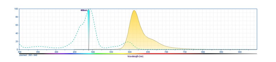
The BD Horizon RealBlue™ 613 (RB613) Dye is part of the BD® family of blue dyes. It is a tandem fluorochrome with an excitation maximum (Ex Max) at 492-nm and an emission maximum (Em Max) at 613-nm as measured using an antibody-dye conjugate. Driven by BD® innovation, RB613 can be used on both spectral and conventional cytometers and is designed to be excited by the Blue laser (488-nm) with reduced excitation by the 561-nm Yellow-Green laser. For conventional instruments equipped with a Blue laser (488-nm), RB613 can be used as an alternative to PE-CF594 and we recommend using an optical filter centered near 610-nm (eg, a 610/20-nm bandpass filter). For spectral instruments equipped with a Blue laser (488-nm), it can be used in conjunction with PE-CF594. RB613 is on average brighter than PE-CF594 off the blue laser.
RB613
492 nm
613 nm
应用
实验应用
Flow cytometry (Routinely Tested)
推荐用量
5 µl/test
反应种属
Human (QC Testing), Rhesus,Cynomolgus,Baboon (Tested in Development)
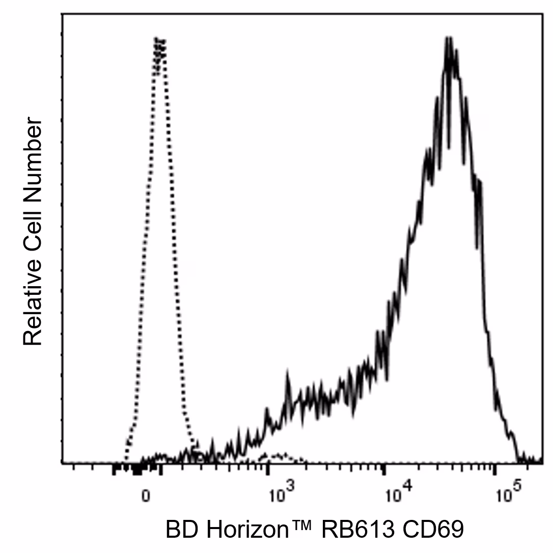
Flow cytometric analysis of CD69 expression on stimulated Human peripheral blood lymphocytes. Human peripheral blood mononuclear cells (PBMC) were stimulated for 24 hours with Phytohemagglutinin (PHA). The cells were then stained with either BD Horizon™ RB613 Mouse IgG1, κ Isotype Control (Cat. No. 571106; dashed line histogram) or BD Horizon™ RB613 Mouse Anti-Human CD69 antibody (Cat. No. 571124/571125; solid line histogram). The fluorescence histogram showing CD69 expression (or Ig Isotype control staining) was derived from gated events with the forward and side light-scatter characteristics of viable activated lymphocytes. Flow cytometry and data analysis were performed using a BD FACSymphony™ A5 SE Cell Analyzer System and FlowJo™ Software.


 全部商品分类


 下载产品说明书
 用小程序,查商品更便捷


 收藏
收藏
 对比
对比 咨询
咨询