


 全部商品分类
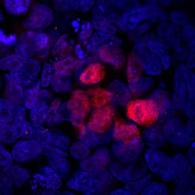


 用小程序,查商品更便捷


Met230-Met435
Accession # O15178
Scientific Data
.") View Larger
View LargerBrachyury in BG01V Human Embryonic Stem Cells. Brachyury was detected in immersion fixed BG01V human embryonic stem cells differentiated into mesoderm using Rabbit Anti-Human Brachyury Monoclonal Antibody (Catalog # MAB20851) at a 10 µg/mL for 3 hours at room temperature. Cells were stained using the NorthernLights™ 557-conjugated Anti-Rabbit IgG Secondary Antibody (red; Catalog # NL004) and counterstained with DAPI (blue). Specific staining was localized to nuclei. View our protocol for Fluorescent ICC Staining of Stem Cells on Coverslips.
Human Brachyury Antibody Summary
Met230-Met435
Accession # O15178
Applications
Please Note: Optimal dilutions should be determined by each laboratory for each application. General Protocols are available in the Technical Information section on our website.
Background: Brachyury
Brachyury is a 435 aa T-box family transcription factor. Human Brachyury shares 90% and 91% aa identity with mouse and rat Brachyury, respectively. It is required in the early determination and differentiation of mesoderm. Additionally, expression of Brachyury has been shown to be upregulated in a number of cancers, and in some cases, correlates with increased aggressiveness.
Preparation and Storage
- 12 months from date of receipt, -20 to -70 °C as supplied.
- 1 month, 2 to 8 °C under sterile conditions after reconstitution.
- 6 months, -20 to -70 °C under sterile conditions after reconstitution.
参考图片
Brachyury in BG01V Human Embryonic Stem Cells. Brachyury was detected in immersion fixed BG01V human embryonic stem cells differentiated into mesoderm using Rabbit Anti-Human Brachyury Monoclonal Antibody (Catalog # MAB20851) at a 1:50 dilution for 3 hours at room temperature. Cells were stained using the NorthernLights™ 557-conjugated Anti-Rabbit IgG Secondary Antibody (red; Catalog # NL004) and counterstained with DAPI (blue). Specific staining was localized to nuclei. View our protocol for Fluorescent ICC Staining of Stem Cells on Coverslips.