


 全部商品分类


Human Exhausted CD8 + T Cell IHC Antibody Sampler Kit

 下载产品说明书 下载SDS
 用小程序,查商品更便捷


 收藏
收藏
 对比
对比 咨询
咨询
The Human Exhausted CD8+ T Cell IHC Antibody Sampler Kit provides an economical means of characterizing the extent of exhaustion in T cells in formalin-fixed, paraffin-embedded tissue samples.
参考图片
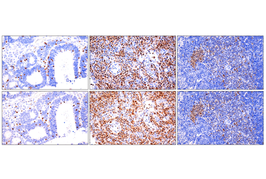
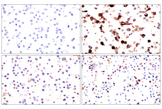
Immunohistochemical analysis of paraffin-embedded human endometrioid adenocarcinoma (left), human T-cell lymphoma (middle), or mouse spleen (right) using Tox/Tox2 (E6I3Q) Rabbit mAb (top) or Tox Rat mAb (bottom). These two antibodies detect independent, unique epitopes on human Tox. The similar staining patterns obtained with both antibodies help to confirm the specificity of the staining.
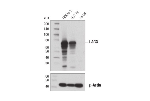
Western blot analysis of extracts from HDLM-2, HuT 78, and Jurkat cells using LAG3 (D2G4O) XP® Rabbit mAb (upper) and β-Actin (D6A8) Rabbit mAb #8457 (lower).
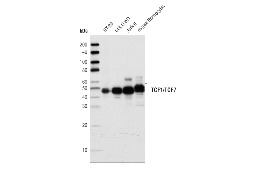
Western blot analysis of total cell lysates from HT29, Colo201, Jurkat and mouse thymocytes using TCF1/TCF7 (C63D9) Rabbit mAb.
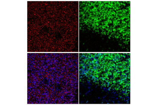
Confocal immunofluorescent analysis of fixed frozen lymph node from wild-type (left) Tcf7GFP flox (right; Jax Strain 030909) mice using TCF1/TCF7 (C63D9) Rabbit mAb #2203 (red) and CD4 (RM4-5) Rat mAb (redFluor™ 710 Conjugate) #75508 (blue). EGFP insertion around Tcf7 exon 2 interferes with expression of long isoforms, but not short isoforms.
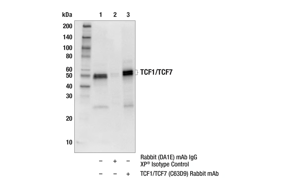
Immunoprecipitation of TCF1/TCF7 protein from Jurkat cell extracts. Lane 1 is 10% input, lane 2 is Rabbit (DA1E) mAb IgG XP® Isotype Control #3900, and lane 3 is TCF1/TCF7 (C63D9) Rabbit mAb. Western blot analysis was performed using TCF1/TCF7 (C63D9) Rabbit mAb. Mouse Anti-rabbit IgG (Conformation Specific) (L27A9) mAb #3678 was used as a secondary antibody.
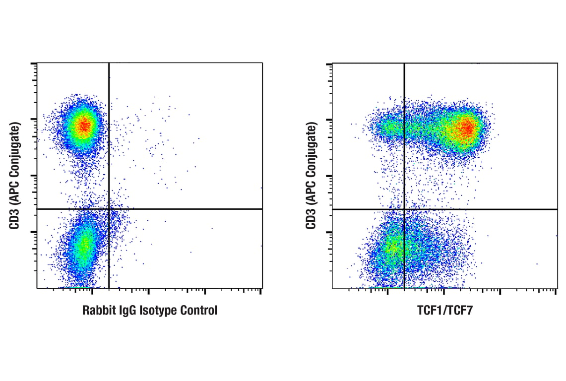
Flow cytometric analysis of human peripheral blood mononuclear cells using TCF1/TCF7 (C63D9) Rabbit mAb (right) or concentration-matched Rabbit (DA1E) mAb IgG XP® Isotype Control #3900 (left), co-stained with CD3 (UCHT1) Mouse mAb (APC Conjugate) #19881. Anti-rabbit IgG (H+L), F(ab')2 Fragment (Alexa Fluor® 488 Conjugate) #4412 was used as a secondary antibody.
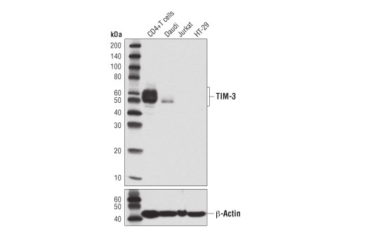
Western blot analysis of extracts from primary human CD4+ T cells and various cell lines using TIM-3 (D5D5R™) XP® Rabbit mAb (upper) or β-Actin (D6A8) Rabbit mAb #8457 (lower). CD4+ T cells were purified from human blood and stimulated for 9 days using beads coated with CD3 and CD28 antibodies in the presence of Human Interleukin-2 (hIL-2) #8907 (6.7 ng/ml).
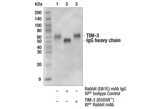
Immunoprecipitation of TIM-3 from RPMI 8226 cell extracts. Lane 1 is 10% input, lane 2 is precipitated with Rabbit (DA1E) mAb IgG XP® Isotype Control #3900, and lane 3 is TIM-3 (D5D5R™) XP® Rabbit mAb, #45208. Western blot was performed using TIM-3 (D5D5R™) XP® Rabbit mAb. Secondary detection was performed using #12291, Protein A (HRP Conjugate).
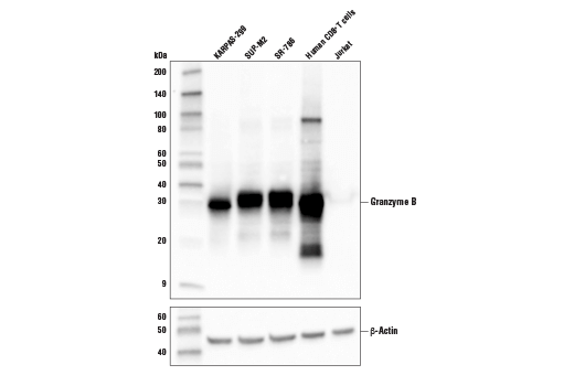
Western blot analysis of extracts from various human cells using Granzyme B (D6E9W) Rabbit mAb (upper) and β-actin (D6A8) Rabbit mAb #8457 (lower). CD8+ T cells were purified from human blood and stimulated for 9 days using beads coated with CD3 and CD28 antibodies in the presence of human interleukin-2 (hIL-2) #8907 (20 ng/ml). KARPAS cell line source: Dr. Abraham Karpas at the University of Cambridge.
Immunohistochemical analysis of paraffin-embedded normal rhesus monkey spleen using Granzyme B (D6E9W) Rabbit mAb.
After the primary antibody is bound to the target protein, a complex with HRP-linked secondary antibody is formed. The LumiGLO® is added and emits light during enzyme catalyzed decomposition.
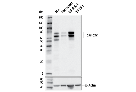
Western blot analysis of extracts from human and mouse cell lines and rat tissue using Tox/Tox2 (E6I3Q) Rabbit mAb (upper) and β-Actin (D6A8) Rabbit mAb #8457 (lower).
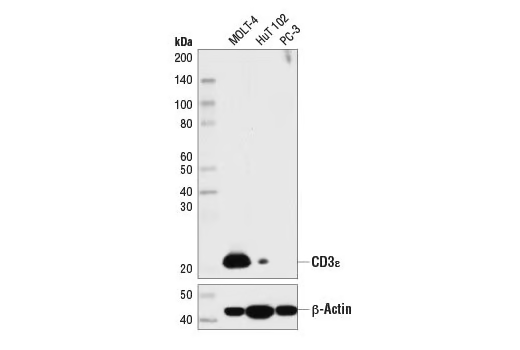
Western blot analysis of extracts from MOLT-4, HuT 102, and PC-3 cells using CD3ε (D7A6E™) XP® Rabbit mAb (upper) or β-Actin (D6A8) Rabbit mAb #8457 (lower).
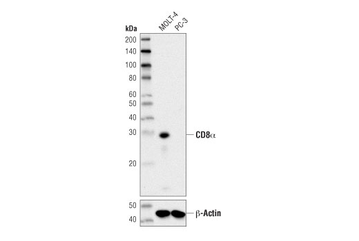
Western blot analysis of extracts from MOLT-4 and PC-3 cells using CD8α (D8A8Y) Rabbit mAb (upper) and β-Actin (D6A8) Rabbit mAb #8457 (lower).
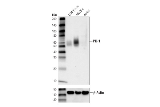
Western blot analysis of extracts from human CD4+ T cells, MOLT-4, and Jurkat cells using PD-1 (Intracellular Domain) (D4W2J) XP® Rabbit mAb (upper), and β-Actin (D6A8) Rabbit mAb #8457 (lower). CD4+ T cells were purified from human blood and stimulated for 9 days using beads coated with CD3 and CD28 antibodies in the presence of human interleukin-2 (hIL-2) #8907 (6.7 ng/ml).
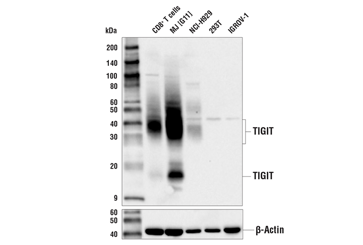
Western blot analysis of extracts from human CD8+ T cells and various human cell lines using TIGIT (E5Y1W) XP® Rabbit mAb (upper) and β-Actin (D6A8) Rabbit mAb #8457 (lower).
Immunohistochemical analysis of paraffin-embedded human breast ductal carcinoma using LAG3 (D2G4O) XP® Rabbit mAb performed on the Leica® Bond™ Rx.
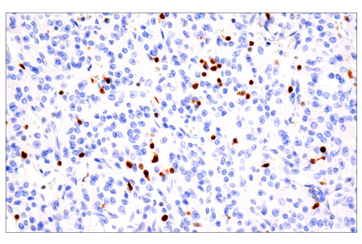
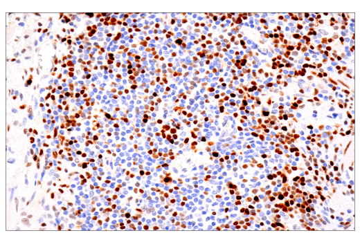
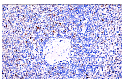
Immunohistochemical analysis of paraffin-embedded human Non-Hodgkin lymphoma using TCF1/TCF7 (C63D9) Rabbit mAb.
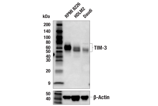
Western blot analysis of extracts from various cell lines using TIM-3 (D5D5R™) XP® Rabbit mAb #45208 (upper) or β-Actin (D6A8) Rabbit mAb #8457 (lower).
Western blot analysis of extracts from CTLL-2, mouse CD8+ T, and C2C12 cells using Granzyme B (D6E9W) Rabbit mAb (upper) and β-actin (D6A8) Rabbit mAb #8457 (lower). CD8+ T cells were purified from mouse spleens and stimulated for 9 days using beads coated with mouse CD3 and CD28 antibodies in the presence of Mouse Interleukin-2 (mIL-2) #5201 (20 ng/ml).
Western blot analysis of extracts from 293T cells, mock transfected (-) or transfected (+) with constructs expressing Myc/DDK-tagged full-length human Tox protein (hTox-Myc/DDK), Myc/DDK-tagged full-length human Tox2 protein (hTox2-Myc/DDK), Myc/DDK-tagged full-length human Tox3 protein (hTox3-Myc/DDK), or Myc/DDK-tagged full-length human Tox4 protein (hTox4-Myc/DDK), using Tox/Tox2 (E6I3Q) Rabbit mAb (upper), Myc-Tag (71D10) Rabbit mAb #2278 (middle), and β-Actin (D6A8) Rabbit mAb #8457 (lower).
Immunohistochemical analysis of paraffin-embedded human mucoepidermoid carcinoma of the larynx using CD3ε (D7A6E™) XP® Rabbit mAb performed on the Leica® BOND™ Rx.
Confocal immunofluorescent analysis of Jurkat cells (left, positive) or Raji cells (right, negative) using CD3ε (D7A6E™) Rabbit mAb (green) and DAPI #4083 (blue).
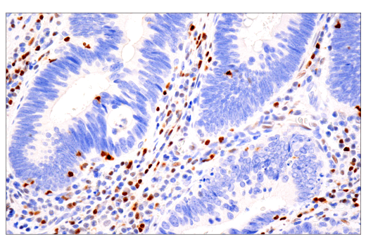
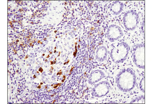
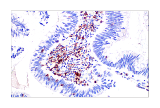
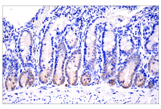
Immunohistochemical analysis of paraffin-embedded human Crohn's diseased colon using CD8a (D8A8Y) Rabbit mAb.
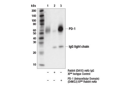
Immunoprecipitation of PD-1 protein from Molt-4 cell extracts. Lane 1 is 10% input, lane 2 is Rabbit (DA1E) mAb IgG XP® Isotype Control #3900, and lane 3 is PD-1 (Intracellular Domain) (D4W2J) XP® Rabbit mAb. Western blot analysis was performed using PD-1 (Intracellular Domain) (D4W2J) XP® Rabbit mAb.
Immunohistochemical analysis of paraffin-embedded HDLM-2 (left) and PC-3 (right) cell pellets on SignalSlide® PD-L1 IHC Controls #13747 using LAG3 (D2G4O) XP® Rabbit mAb.
Immunohistochemical analysis of paraffin-embedded human lung carcinoma using TCF1/TCF7 (C63D9) Rabbit mAb in the presence of control peptide (left) or TCF1/TCF7 blocking peptide #1007 (right).
Immunohistochemical analysis of paraffin-embedded renal clear cell carcinoma using TIM-3 (D5D5R™) XP® Rabbit mAb performed on the Leica® BOND™ Rx.
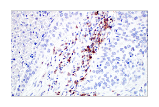
Immunohistochemical analysis of paraffin-embedded human colon adenocarinoma using Granzyme B (D6E9W) Rabbit mAb performed on the Leica® BOND™ Rx.
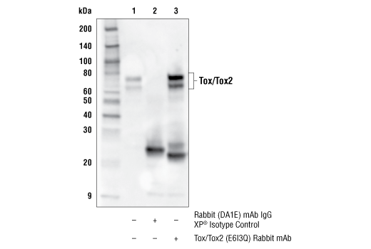
Immunoprecipitation of Tox/Tox2 protein from SU-DHL-4 cell extracts. Lane 1 is 10% input, lane 2 is Rabbit (DA1E) mAb IgG XP® Isotype Control #3900, and lane 3 is Tox/Tox2 (E6I3Q) Rabbit mAb. Western blot analysis was performed using Tox/Tox2 (E6I3Q) Rabbit mAb.
Immunohistochemical analysis of paraffin-embedded human endometrioid adenocarcinoma using CD3ε (D7A6E™) XP® Rabbit mAb performed on the Leica® BOND™ Rx.
Immunohistochemical analysis of paraffin-embedded human lung carcinoma using CD8a (D8A8Y) Rabbit mAb.
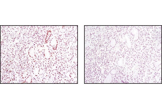
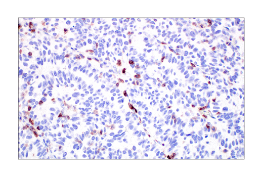
Immunohistochemical analysis of paraffin-embedded human infiltrating papillary carcinoma of the breast using PD-1 (Intracellular Domain) (D4W2J) XP® Rabbit mAb performed on the Leica® BOND™ Rx.
Immunohistochemical analysis of paraffin-embedded 293T cell pellets, control (left-top) or TIGIT-transfected (right-top), MJ [G11] cell pellet (left-bottom, positive), and purified CD8+ human peripheral blood mononuclear cell pellet (right-bottom, positive), using TIGIT (E5Y1W) XP® Rabbit mAb. CD8+ T cells were purified from human blood and stimulated for 7 days using beads coated with CD3 and CD28 antibodies in the presence of Human Interleukin-2 (hIL-2) #8907 (7 ng/mL).
Immunohistochemical analysis of paraffin-embedded human colitis using LAG3 (D2G4O) XP® Rabbit mAb.
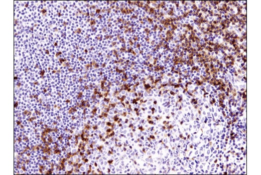
Immunohistochemical analysis of paraffin-embedded human tonsil using TCF1/TCF7 (C63D9) Rabbit mAb.
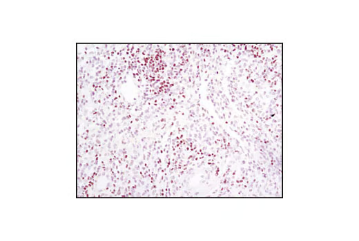
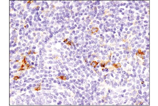
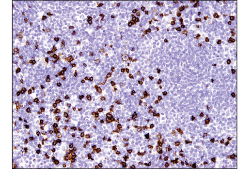
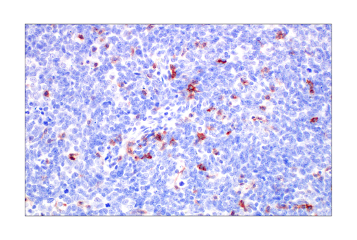
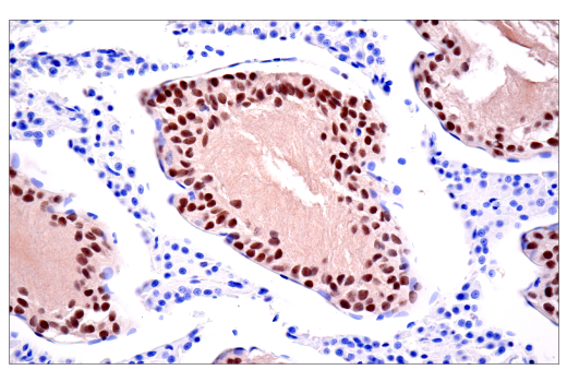
Immunohistochemical analysis of paraffin-embedded human tonsil using TIM-3 (D5D5R™) XP® Rabbit mAb.
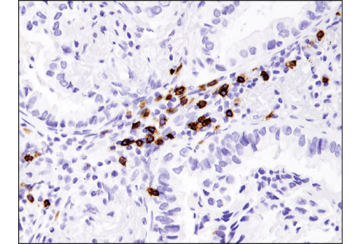
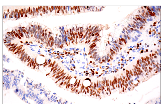
Immunohistochemical analysis of paraffin-embedded human colon carcinoma using Tox/Tox2 (E6I3Q) Rabbit mAb performed on the Leica® BOND™ Rx.
Immunohistochemical analysis of paraffin-embedded human tonsil using CD3ε (D7A6E™) XP® Rabbit mAb.
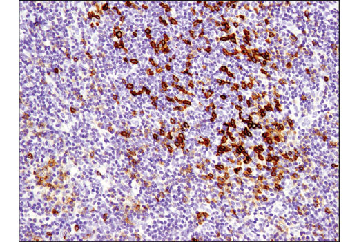
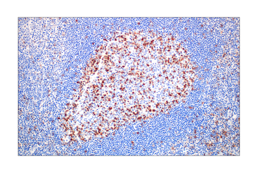
Immunohistochemical analysis of paraffin-embedded human lymphoma using CD8a (D8A8Y) Rabbit mAb.
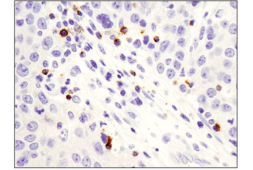
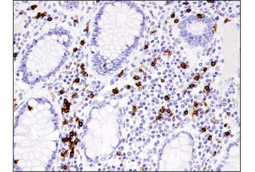
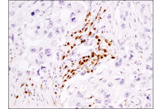
Immunohistochemical analysis of paraffin-embedded human colon carcinoma using PD-1 (Intracellular Domain) (D4W2J) XP® Rabbit mAb.
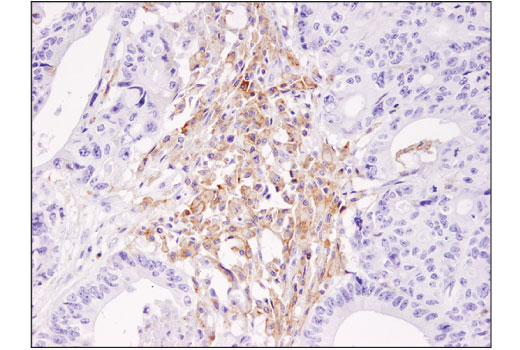
Immunohistochemical analysis of paraffin-embedded human urothelial carcinoma using TIGIT (E5Y1W) XP® Rabbit mAb.
Immunohistochemical analysis of paraffin-embedded human tonsil using LAG3 (D2G4O) XP® Rabbit mAb.
Confocal immunofluorescent analysis of DLD-1 cells using TCF1/TCF7 (C63D9) Rabbit mAb (green). Actin filaments have been labeled with Alexa Fluor® 555 phalloidin (red).
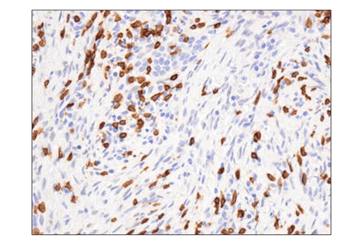
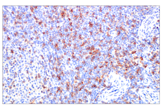
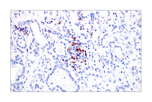
Immunohistochemical analysis of paraffin-embedded human colon carcinoma using TIM-3 (D5D5R™) XP® Rabbit mAb.
Immunohistochemical analysis of paraffin-embedded human papillary carcinoma of the breast using Granzyme B (D6E9W) Rabbit mAb.
Immunohistochemical analysis of paraffin-embedded human colon carcinoma using Tox/Tox2 (E6I3Q) Rabbit mAb performed on the Leica® BOND™ Rx.
Immunohistochemical analysis of paraffin-embedded human breast carcinoma using CD3ε (D7A6E™) XP® Rabbit mAb.
Immunohistochemical analysis of paraffin-embedded normal rhesus monkey spleen using CD8α (D8A8Y) Rabbit mAb.
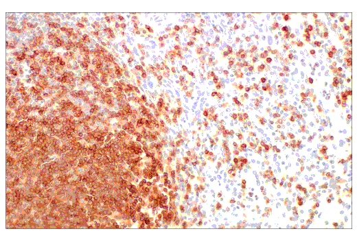
Immunohistochemical analysis of paraffin-embedded human B-cell non-Hodgkin lymphoma using PD-1 (Intracellular Domain) (D4W2J) XP® Rabbit mAb.
Immunohistochemical analysis of paraffin-embedded human colon adenocarcinoma using TIGIT (E5Y1W) XP® Rabbit mAb.
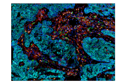
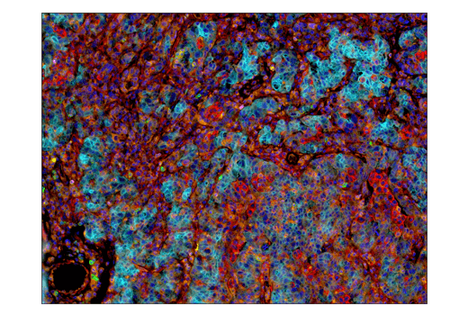
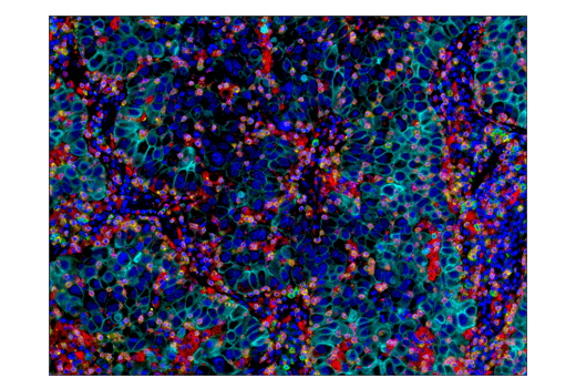
Multiplex immunohistochemical analysis of paraffin-embedded human breast carcinoma usng LAG3 (D2G4O) XP® rabbit mAb (magenta), PD-1 (D4W2J) XP® rabbit mAb #86163 (green), PD-L1 (E1L3N®) XP® rabbit mAb #13684 (red), TIM-3 (D5D5R™) XP® rabbit mAb #45208 (yellow), CD8α (C8/144B) mouse mAb #70306 (orange), and Pan-keratin (C11) mouse mAb #4545 (cyan).
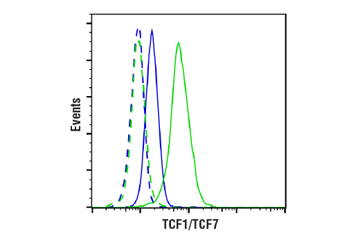
Flow cytometric analysis of A20 cells (blue) and EL-4 cells (green) using TCF1/TCF7 (C63D9) Rabbit mAb (solid lines) or a concentration-matched Rabbit (DA1E) mAb IgG XP® Isotype Control #3900 (dashed lines). Anti-rabbit IgG (H+L), F(ab')2 Fragment (Alexa Fluor® 488 Conjugate) #4412 was used as a secondary antibody.
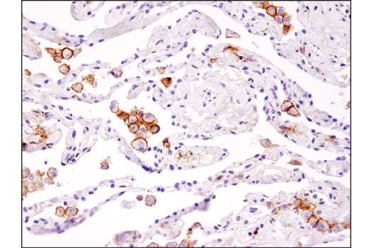
Immunohistochemical analysis of paraffin-embedded human lung carcinoma using TIM-3 (D5D5R™) XP® Rabbit mAb. Note staining of alveolar macrophages.
Immunohistochemical analysis of paraffin-embedded human renal cell carcinoma using Tox/Tox2 (E6I3Q) Rabbit mAb performed on the Leica® BOND™ Rx.
Immunohistochemical analysis of paraffin-embedded human non-Hodgkin lymphoma using CD3ε (D7A6E™) XP® Rabbit mAb.
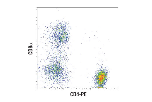
Flow cytometric analysis of fixed and permeabilized human whole blood using CD8α (D8A8Y) Rabbit mAb co-stained with CD4-PE, showing a distinct CD8α positive population with no cross reactivity to CD4 positive cells. Anti-rabbit IgG (H+L), F(ab')2 Fragment (Alexa Fluor® 647 Conjugate) #4414 was used as a secondary antibody.
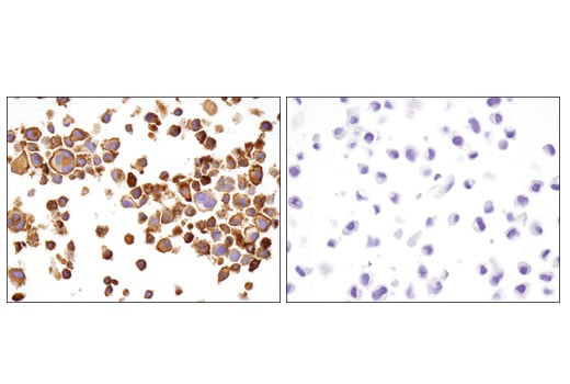
Immunohistochemical analysis of paraffin-embedded 293 cell pellets, control (left) or PD-1 transfected (right), using PD-1 (Intracellular Domain) (D4W2J) XP® Rabbit mAb.
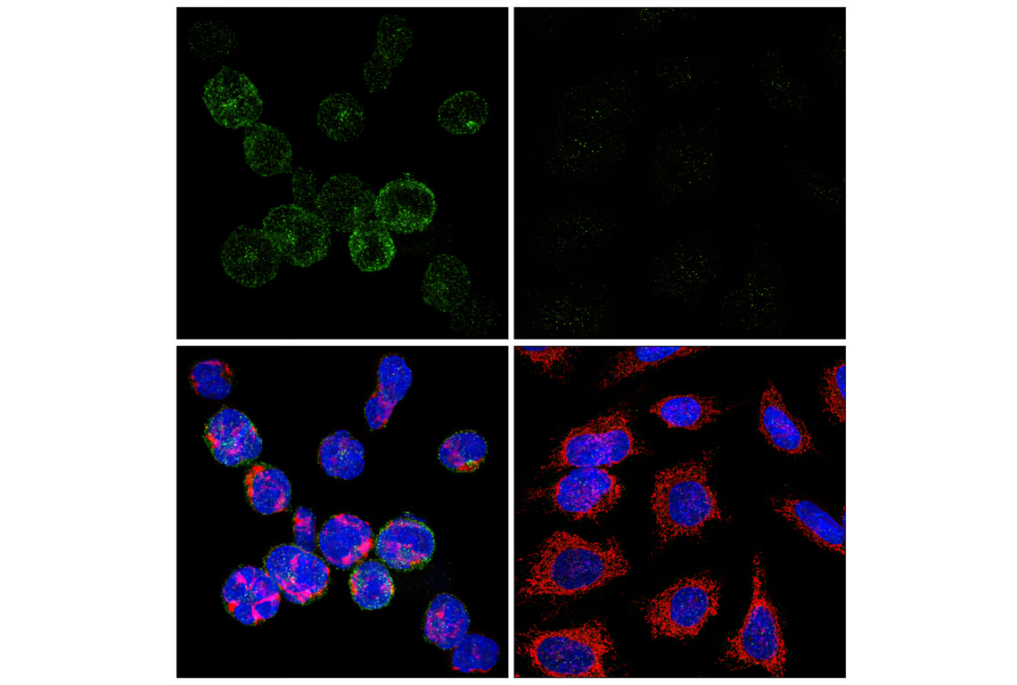
Confocal immunofluorescent analysis of MOLT-4 cells (left, positive) or HeLa cells (right, negative) using PD-1 (Intracellular Domain) (D4W2J) XP® Rabbit mAb (green), Cytochrome c (6H2.B4) Mouse mAb #12963 (red), and DAPI #4083 (blue).
Immunohistochemical analysis of paraffin-embedded human esophageal carcinoma using TIGIT (E5Y1W) XP® Rabbit mAb.
Multiplex immunohistochemical analysis of paraffin-embedded human lung adenocarcinoma using LAG3 (D2G4O) XP® rabbit mAb (orange), PD-1 (EH33) mouse mAb #43248 (green), CD8α (C8/144B) mouse mAb #70306 (magenta), CD68 (D4B9C) XP® rabbit mAb #76437 (red), Pan-keratin (C11) mouse mAb #4545 (cyan), and TIM-3 (D5D5R™) XP® rabbit mAb #45208 (yellow).
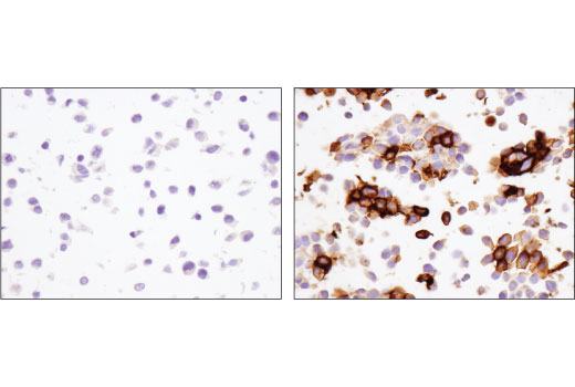
Immunohistochemical analysis of paraffin-embedded cell pellets, primary CD4+ T cells (left) and HT-29 cells (right), using TIM-3 (D5D5R™) XP® Rabbit mAb. CD4+ T cells were purified from human blood and stimulated for 7 days using beads coated with CD3 and CD28 antibodies in the presence of Human Interleukin-2 (hIL-2) #8907 (6.7 ng/ml).
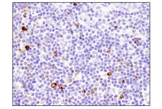
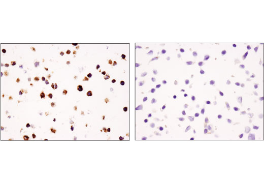
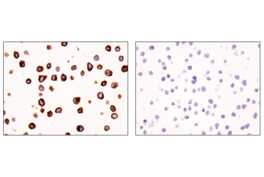
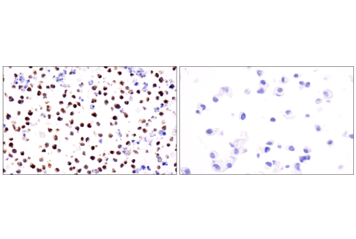
Immunohistochemical analysis of paraffin-embedded KARPAS-299 cell pellet (left, positive) or Jurkat cell pellet (right, negative) using Granzyme B (D6E9W) Rabbit mAb. KARPAS cell line source: Dr. Abraham Karpas at the University of Cambridge.
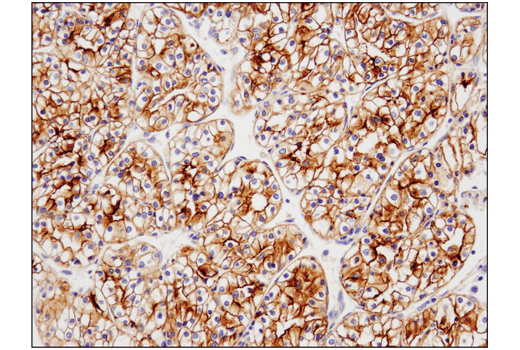
Immunohistochemical analysis of paraffin-embedded human gastric adenocarcinoma using Tox/Tox2 (E6I3Q) Rabbit mAb performed on the Leica® BOND™ Rx.
Immunohistochemical analysis of paraffin-embedded normal cynomolgus monkey spleen using CD3ε (D7A6E™) XP® Rabbit mAb.
Multiplex immunohistochemical analysis of paraffin-embedded human breast carcinoma usng PD-1 (Intracellular Domain) (D4W2J) XP® rabbit mAb (green), PD-L1 (E1L3N®) XP® rabbit mAb #13684 (red), LAG3 (D2G4O™) XP® rabbit mAb #15372 (magenta), TIM-3 (D5D5R™) XP® rabbit mAb #45208 (yellow), CD8α (C8/144B) mouse mAb #70306 (orange), and Pan-keratin (C11) mouse mAb #4545 (cyan).
Immunohistochemical analysis of paraffin-embedded human squamous cell carcinoma of the tonsil using TIGIT (E5Y1W) XP® Rabbit mAb.
Multiplex immunohistochemical analysis of paraffin-embedded human ovarian carcinoma using LAG3 (D2G4O) XP® rabbit mAb (magenta), PD-1 (D4W2J) XP® rabbit mAb #86163 (green), B7-H3 (D9M2L) XP® rabbit mAb #14058 (red), B7-H4 (D1M8I) XP® rabbit mAb (cyan), TIM-3 (D5D5R™) XP® rabbit mAb #45208 (yellow), and VISTA (D1L2G™) XP® rabbit mAb #64953 (orange).
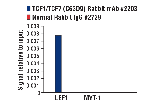
Chromatin immunoprecipitations were performed with cross-linked chromatin from mouse thymocytes and either TCF1/TCF7 (C63D9) Rabbit mAb or Normal Rabbit IgG #2729 using SimpleChIP® Plus Enzymatic Chromatin IP Kit (Magnetic Beads) #9005. The enriched DNA was quantified by real-time PCR using SimpleChIP® Mouse LEF1 Upstream Primers #80993 and SimpleChIP® Mouse MYT-1 Promoter Primers #8985. The amount of immunoprecipitated DNA in each sample is represented as signal relative to the total amount of input chromatin, which is equivalent to one.
Multiplex immunohistochemical analysis of paraffin-embedded human breast carcinoma usng TIM-3 (D5D5R™) XP® rabbit mAb (yellow), PD-1 (D4W2J) XP® rabbit mAb #86163 (green), PD-L1 (E1L3N®) XP® rabbit mAb #13684 (red), LAG3 (D2G4O™) XP® rabbit mAb #15372 (magenta), CD8α (C8/144B) mouse mAb #70306 (orange), and Pan-keratin (C11) mouse mAb #4545 (cyan).
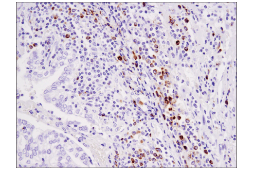
Immunohistochemical analysis of paraffin-embedded human colon carcinoma using Granzyme B (D6E9W) Rabbit mAb.
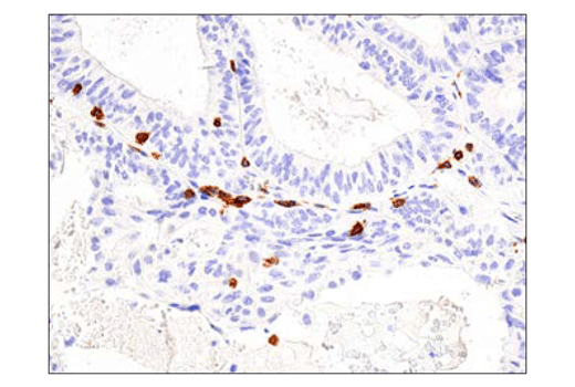
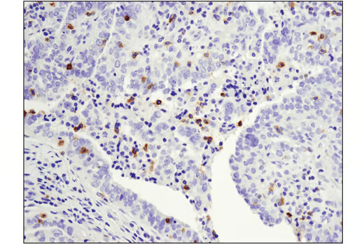
Immunohistochemical analysis of paraffin-embedded normal human spleen using Tox/Tox2 (E6I3Q) Rabbit mAb.
Immunohistochemical analysis of paraffin-embedded MOLT-4 (left) and PC-3 (right) cell pellets using CD3ε (D7A6E™) XP® Rabbit mAb.
Multiplex immunohistochemical analysis of paraffin-embedded human ovarian carcinoma using PD-1 (Intracellular Domain) (D4W2J) XP® rabbit mAb (green), B7-H3 (D9M2L) XP® rabbit mAb #14058 (red), B7-H4 (D1M8I) XP® rabbit mAb (cyan), LAG3 (D2G4O™) XP® rabbit mAb #15372 (magenta), TIM-3 (D5D5R™) XP® rabbit mAb #45208 (yellow), and VISTA (D1L2G™) XP® rabbit mAb #64953 (orange).
Immunohistochemical analysis of paraffin-embedded human non-small cell lung carcinoma using TIGIT (E5Y1W) XP® Rabbit mAb.
Multiplex immunohistochemical analysis of paraffin-embedded human lung adenocarcinoma using TIM-3 (D5D5R™) XP® rabbit mAb (yellow), PD-1 (EH33) mouse mAb #43248 (green), CD8α (C8/144B) mouse mAb #70306 (magenta), CD68 (D4B9C) XP® rabbit mAb #76437 (red), Pan-keratin (C11) mouse mAb #4545 (cyan), and LAG3 (D2G4O™) XP® rabbit mAb #15372 (orange).
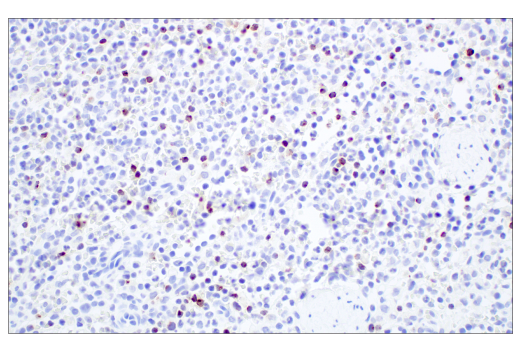
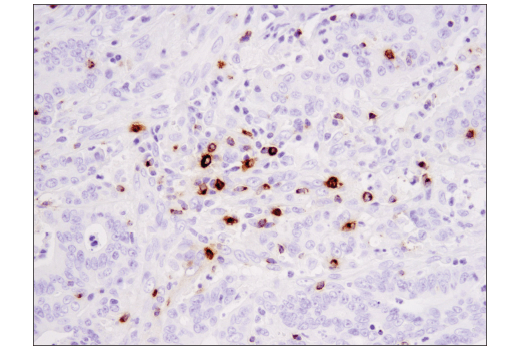
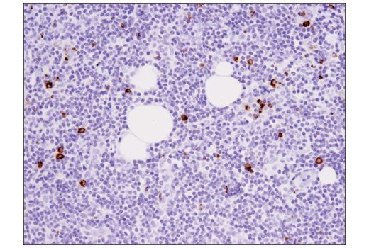
Immunohistochemical analysis of paraffin-embedded human non-Hodgkin lymphoma using Granzyme B (D6E9W) Rabbit mAb.
Immunohistochemical analysis of paraffin-embedded rat testis using Tox/Tox2 (E6I3Q) Rabbit mAb.
Immunohistochemical analysis of paraffin-embedded human B-cell non-Hodgkin lymphoma using TIGIT (E5Y1W) XP® Rabbit mAb (left) compared to concentration-matched Rabbit (DA1E) mAb IgG XP® Isotype Control #3900 (right).
Multiplex immunohistochemical analysis of paraffin-embedded human ovarian carcinoma using TIM-3 (D5D5R™) XP® rabbit mAb (yellow), PD-1 (D4W2J) XP® rabbit mAb #86163 (green), B7-H3 (D9M2L) XP® rabbit mAb #14058 (red), B7-H4 (D1M8I) XP® rabbit mAb (cyan), LAG3 (D2G4O™) XP® rabbit mAb #15372 (magenta), and VISTA (D1L2G™) XP® rabbit mAb #64953 (orange).
Immunohistochemical analysis of paraffin-embedded rat spleen using Tox/Tox2 (E6I3Q) Rabbit mAb.
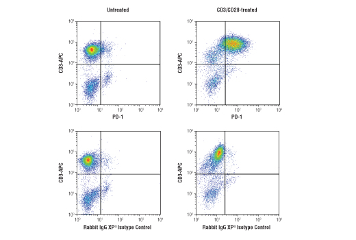
Flow cytometric analysis of fixed and permeabilized human peripheral blood mononuclear cells, untreated (left column) or treated with anti-CD3 (10ug/ml, 72hr) and anti-CD28 (5ug/ml, 72 hr; right column), using PD-1 (Intracellular Domain) (D4W2J) XP® Rabbit mAb #86163 (top row) or concentration-matched Rabbit (DA1E) mAb IgG XP® Isotype Control #3900 (bottom row), and co-stained with CD3 (UCHT1) Mouse mAb (APC Conjugate) #19881. Anti-rabbit IgG (H+L), F(ab')2 Fragment (Alexa Fluor® 488 Conjugate) #4412 was used as a secondary antibody.
Immunohistochemical analysis of paraffin-embedded human prostate adenocarcinoma using TIGIT (E5Y1W) XP® Rabbit mAb.
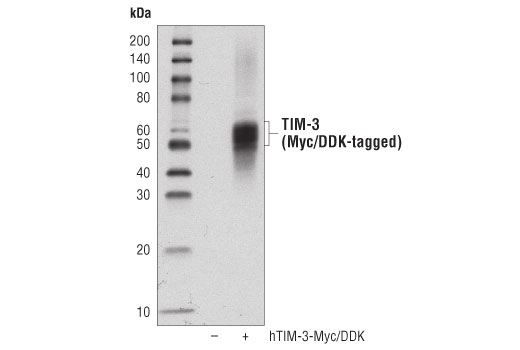
Western blot analysis of extracts from 293T cells, untransfected (-) or transfected with a construct expressing full-length human Myc/DDK-tagged TIM-3 (hTIM-3-Myc/DDK; +), using TIM-3 (D5D5R™) XP® Rabbit mAb.
Immunohistochemical analysis of paraffin-embedded rat small intestine using Tox/Tox2 (E6I3Q) Rabbit mAb.
Immunohistochemical analysis of paraffin-embedded human tonsil using TIGIT (E5Y1W) XP® Rabbit mAb.
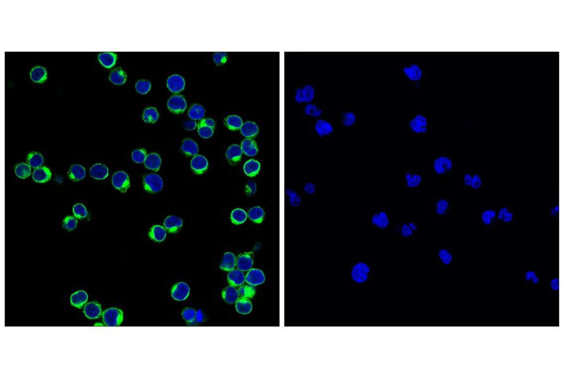
Flow cytometric analysis of Jurkat cells (blue) and primary CD4+ T cells (green) using TIM-3 (D5D5R™) XP® Rabbit mAb (solid lines) or a concentration-matched Rabbit (DA1E) mAb IgG XP® Isotype Control #3900 (dashed lines). Anti-rabbit IgG (H+L), F(ab')2 fragment (Alexa Fluor® 488 Conjugate) #4412 was used as a secondary antibody. CD4+ T cells were purified from human blood and stimulated for 9 days using beads coated with CD3 and CD28 antibodies in the presence of Human Interleukin-2 (hIL-2) #8907 (6.7 ng/ml).
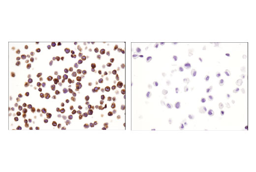
Immunohistochemical analysis of paraffin-embedded SU-DHL-4 cell pellet (left, positive) or ZR-75-1 cell pellet (right, negative) using Tox/Tox2 (E6I3Q) Rabbit mAb.
Confocal immunofluorescent analysis of human CD8+ T cells using TIGIT (E5Y1W) XP® Rabbit mAb (green) and CD8α (RPA-T8) Mouse mAb (FITC Conjugate) #55397 (red pseudocolor). Samples were mounted in ProLong® Gold Antifade Reagent with DAPI #8961 (blue). CD8+ T cells were purified from human blood and stimulated for 7 days using beads coated with CD3 and CD28 antibodies in the presence of Human Interleukin-2 (hIL-2) #8907 (20 ng/mL).
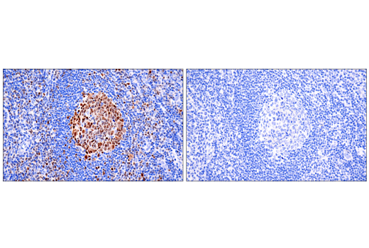
Immunohistochemical analysis of paraffin-embedded human tonsil using Tox/Tox2 (E6I3Q) Rabbit mAb (left) compared to concentration-matched Rabbit (DA1E) mAb IgG XP® Isotype Control #3900 (right).
Confocal immunofluorescent analysis of MJ [G11] cells (left, positive) and IGROV-1 cells (right, negative) using TIGIT (E5Y1W) XP® Rabbit mAb (green). Samples were mounted in ProLong® Gold Antifade Reagent with DAPI #8961 (blue).