


 全部商品分类
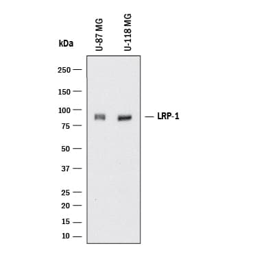


 用小程序,查商品更便捷


Scientific Data
 View Larger
Detection of Human LRP‑1 Cluster II by Western Blot. Western blot shows lysates of U‑87 MG human glioblastoma/astrocytoma cell line and U‑118‑MG human glioblastoma/astrocytoma cell line. PVDF membrane was probed with 2 µg/mL of Mouse Anti-Human LRP‑1 Cluster II Monoclonal Antibody (Catalog # MAB2368) followed by HRP-conjugated Anti-Mouse IgG Secondary Antibody (HAF018). A specific band was detected for LRP‑1 Cluster II at approximately 85 kDa (as indicated). This experiment was conducted under reducing conditions and using Western Blot Buffer Group 1.
 View Larger
LRP‑1 Cluster II in Human Brain (Cortex). LRP‑1 Cluster II was detected in immersion fixed paraffin-embedded sections of human brain (cortex) using Mouse Anti-Human LRP‑1 Cluster II Monoclonal Antibody (Catalog # MAB2368) at 5 µg/mL for 1 hour at room temperature followed by incubation with the Anti-Mouse IgG VisUCyte™ HRP Polymer Antibody (VC001). Before incubation with the primary antibody, tissue was subjected to heat-induced epitope retrieval using Antigen Retrieval Reagent-Basic (CTS013). Tissue was stained using DAB (brown) and counterstained with hematoxylin (blue). Specific staining was localized to cytoplasm in neurons. Staining was performed using our protocol for IHC Staining with VisUCyte HRP Polymer Detection Reagents.
Human LRP-1 Cluster II Antibody Summary
Gln4449-Ala4544
Accession # Q07954
Applications
Please Note: Optimal dilutions should be determined by each laboratory for each application. General Protocols are available in the Technical Information section on our website.
Background: LRP-1 Cluster II
LRP-1 (low-density lipoprotein receptor-related protein 1) is a large type I transmembrane protein belonging to the LDL receptor superfamily. The extracellular domain of LRP-1 is organized in four clusters of ligand-binding repeats that recognizes at least 30 different ligands. The amino acid sequence of human and mouse LRP-1 cluster II share 100% identity. LRP-1 is an endocytic receptor involved in endocytosis and in phagocytosis of apoptotic cells. It plays a crucial role in early embryonic development.
Preparation and Storage
- 12 months from date of receipt, -20 to -70 °C as supplied.
- 1 month, 2 to 8 °C under sterile conditions after reconstitution.
- 6 months, -20 to -70 °C under sterile conditions after reconstitution.