


 全部商品分类
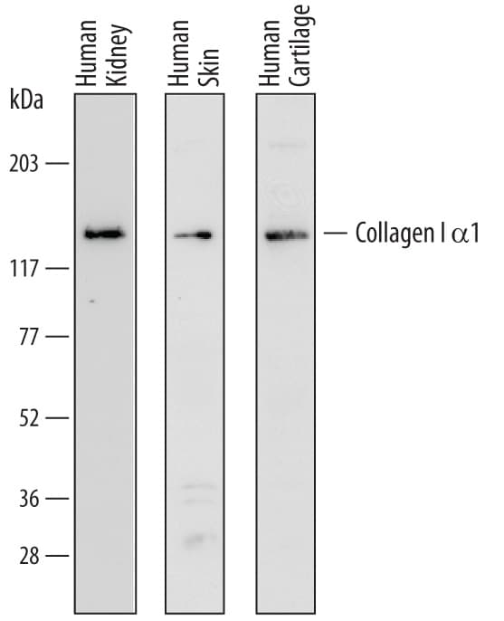


hProCOL1A1 PAb (100 ug)

 下载产品说明书 下载SDS
 用小程序,查商品更便捷


 收藏
收藏
 对比
对比 咨询
咨询Immunocytochemistry(5-15 µg/mL)

Gln23-Lys277, Gly1094-Leu1464
Accession # P02452
Scientific Data
 View Larger
Detection of Human Pro-Collagen I alpha 1 by Western Blot. Western blot shows lysates of human kidney tissue, human skin tissue, and human cartilage tissue. PVDF membrane was probed with 1 µg/mL of Sheep Anti-Human Pro-Collagen I alpha 1 Antigen Affinity-purified Polyclonal Antibody (Catalog # AF6220) followed by HRP-conjugated Anti-Sheep IgG Secondary Antibody (Catalog # HAF016). A specific band was detected for Pro-Collagen I alpha 1 at approximately 140 kDa (as indicated). This experiment was conducted under reducing conditions and using Immunoblot Buffer Group 1.
 View Larger
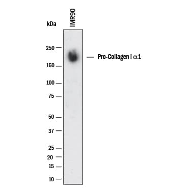
Detection of Human Collagen I by Western Blot. Western blot shows lysates of IMR‑90 human lung fibroblast cell line. PVDF membrane was probed with 1 µg/mL of Sheep Anti-Human Pro Collagen I alpha 1 Antigen Affinity-purified Polyclonal Antibody (Catalog # AF6220) followed by HRP-conjugated Anti-Sheep IgG Secondary Antibody (HAF016). A specific band was detected for Collagen I at approximately 170 kDa (as indicated). This experiment was conducted under reducing conditions and using Western Blot Buffer Group 1.
.") View Larger
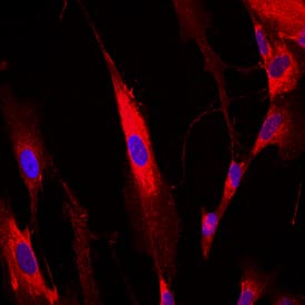
View LargerPro-Collagen I alpha 1 in IMR‑90 Human Cell Line. Pro-Collagen I alpha 1 was detected in immersion fixed IMR-90 human lung fibroblast cell line using Sheep Anti-Human Pro-Collagen I alpha 1 Antigen Affinity-purified Polyclonal Antibody (Catalog # AF6220) at 10 µg/mL for 3 hours at room temperature. Cells were stained using the NorthernLights™ 557-conjugated Anti-Sheep IgG Secondary Antibody (red; Catalog # NL010) and counterstained with DAPI (blue). View our protocol for Fluorescent ICC Staining of Cells on Coverslips.
 View Larger
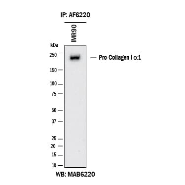
Detection of Human N-Pro Collagen I by Immunoprecipitation. Human N-Pro Collagen I was immunoprecipitated from 500 μg of IMR‑90 human lung fibroblast cell line lysates with 5 μg of Sheep Anti-Human N-Pro Collagen I Antigen Affinity-purified Polyclonal Antibody (Catalog # AF6220). The N-Pro Collagen I-antibody complexes were absorbed using Protein A or Protein G. Immunoprecipitated human N-Pro Collagen I was detected by Western blot using 1 μg/mL of Mouse Anti-Human N-Pro Collagen I Monoclonal Antibody (MAB6220) under reducing conditions and using Western Blot Buffer Group 1.
 View Larger
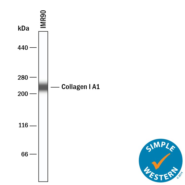
Detection of Human Collagen I alpha 1 by Simple WesternTM. Simple Western lane view shows lysates of IMR‑90 human lung fibroblast cell line, loaded at 0.2 mg/mL. A specific band was detected for Collagen I alpha 1 at approximately 232 kDa (as indicated) using 10 µg/mL of Sheep Anti-Human Pro Collagen I alpha 1 Antigen Affinity-purified Polyclonal Antibody (Catalog # AF6220) followed by 1:50 dilution of HRP-conjugated Anti-Sheep IgG Secondary Antibody (HAF016). This experiment was conducted under reducing conditions and using the 66-440 kDa separation system.
Human Pro Collagen I alpha 1 Antibody Summary
Gln23-Lys277, Gly1094-Leu1464
Accession # P02452
Applications
Please Note: Optimal dilutions should be determined by each laboratory for each application. General Protocols are available in the Technical Information section on our website.
Immunocytochemistry(5-15 µg/mL)
Background: Collagen I alpha 1
Type I collagen is the most abundant structural protein of connective tissues such as skin, bone and tendon. It is synthesized as a procollagen molecule which is characterized by a 300 nm triple helical domain flanked by globular N- and C-terminal propeptides (1). The triple helical domain contains Gly-Xaa-Yaa triplets where Xaa and Yaa are frequently proline and hydroxyproline, respectively. The non-helical propeptides are removed by procollagen N- and C-proteinase activities so that the mature triple helices can self-assemble into collagen fibrils that provide tensile strength to tissues (1). Type I collagen is a heterotrimer that consists of two alpha 1(I) chains and one alpha 2(I) chain, although homotrimers consisting of three identical alpha 1(I) chains have also been described (2). This recombinant mini pro-alpha 1(I) collagen consists of a shortened alpha 1(I) chain with following domain structure from N- to C-terminus: N-propeptide, N‑telopeptide, the 33 most N-terminal Gly-Xaa-Yaa repeats, the 33 most C-terminal Gly-Xaa-Yaa repeats, C-telopeptide and C-propeptide. The preparation contains a mixture of the full-length molecule, pN collagen I( alpha 1) and the C-terminal propeptide. This truncated pro-alpha 1(I) collagen is a substrate for procollagen N-proteinase and procollagen C-proteinase.
- Canty, E.G. et al. (2005) J. Cell Sci. 118:1341.
- Han, S. et al. (2008) J. Mol. Biol. 383:122.
Preparation and Storage
- 12 months from date of receipt, -20 to -70 °C as supplied.
- 1 month, 2 to 8 °C under sterile conditions after reconstitution.
- 6 months, -20 to -70 °C under sterile conditions after reconstitution.
参考图片
Detection of Human Collagen I alpha 1 by Western Blot. Western blot shows lysates of human kidney tissue, human skin tissue, and human cartilage tissue. PVDF membrane was probed with 1 µg/mL of Sheep Anti-Human Collagen I alpha 1 Antigen Affinity-purified Polyclonal Antibody (Catalog # AF6220) followed by HRP-conjugated Anti-Sheep IgG Secondary Antibody (Catalog # HAF016). A specific band was detected for Collagen I alpha 1 at approximately 140 kDa (as indicated). This experiment was conducted under reducing conditions and using Immunoblot Buffer Group 1.
Collagen I alpha 1 in IMR‑90 Human Cell Line. Collagen I alpha 1 was detected in immersion fixed IMR‑90 human lung fibroblast cell line using Sheep Anti-Human Collagen I alpha 1 Antigen Affinity-purified Polyclonal Antibody (Catalog # AF6220) at 10 µg/mL for 3 hours at room temperature. Cells were stained using the NorthernLights™ 557-conjugated Anti-Sheep IgG Secondary Antibody (red; Catalog # NL010) and counterstained with DAPI (blue). View our protocol for Fluorescent ICC Staining of Cells on Coverslips.