


1/66

品牌: CST
 下载产品说明书
 用小程序,查商品更便捷


 收藏
收藏
 对比
对比 咨询
咨询
产品介绍
产品信息
抗原名称
Human Reactive Cell Death and Autophagy

来源纯化
Monoclonal antibodies are produced by immunizing animals with synthetic peptides corresponding to residues surrounding Asp175 of human caspase-3, Asp214 of human PARP, Asp275 of human Gasdermin D, Asp116 of human IL-1β, Pro220 of human SQSTM1/p62, residues near the amino terminus of human LC3B, and synthetic phosphopeptides corresponding to Ser166 of human RIP, Ser227 of human RIP3, and Ser358 of human MLKL.
简单描述
Antibody Sampler Kit for studying RIPK1 (Ser166) phosphate/IL1 beta (Asp116) cleaved/SQSTM1/RIPK3 (Ser227) phosphate/gasdermin D (Asp275) cleaved/MLKL (Thr357/Ser358) phosphate/Casp3 (Asp175) cleaved/LC3B/PARP1 (Asp214, 89 kD) cleaved in the research area.
研究领域
癌症,细胞生物学,纤维化,免疫学和肿瘤学,代谢,神经科学
应用
目标/特异性
Specificity/Sensitivity
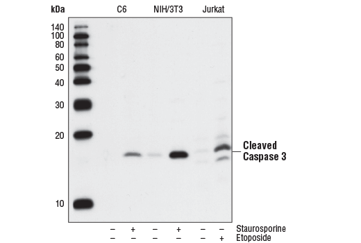
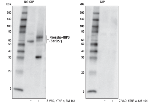
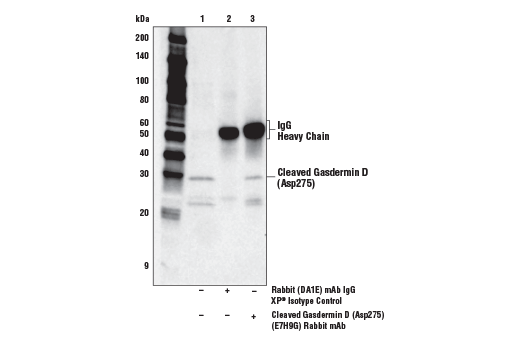
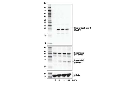
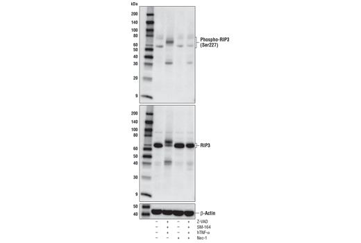
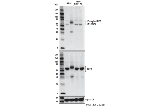
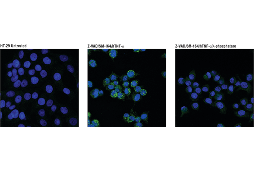
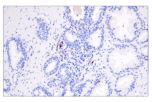
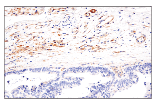
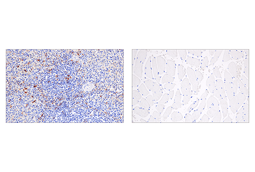
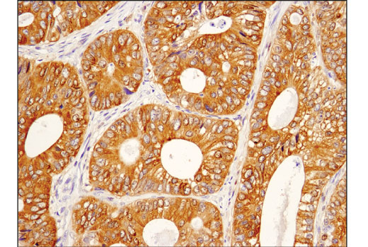
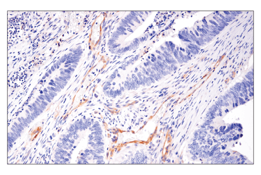
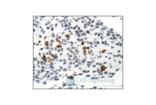
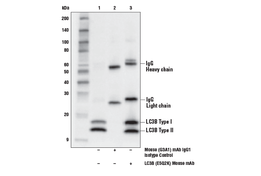
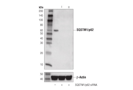
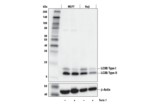
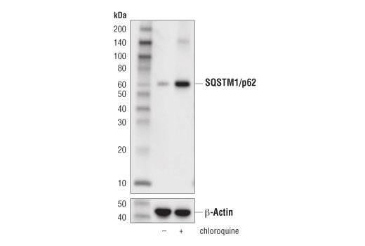
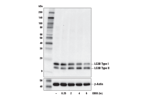
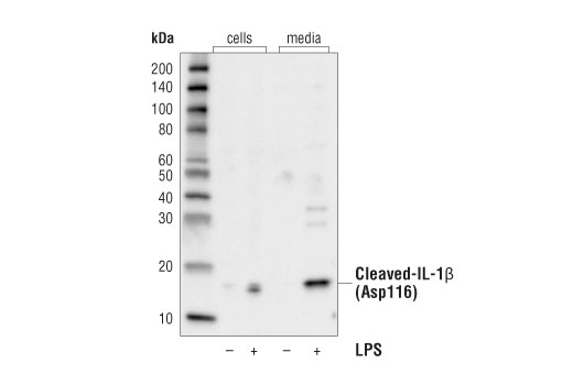
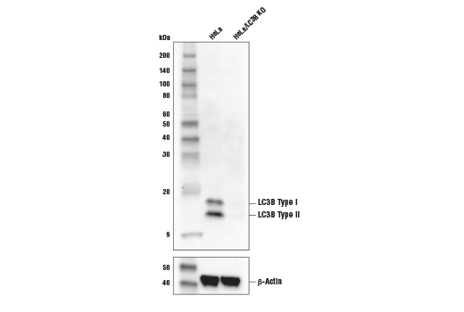
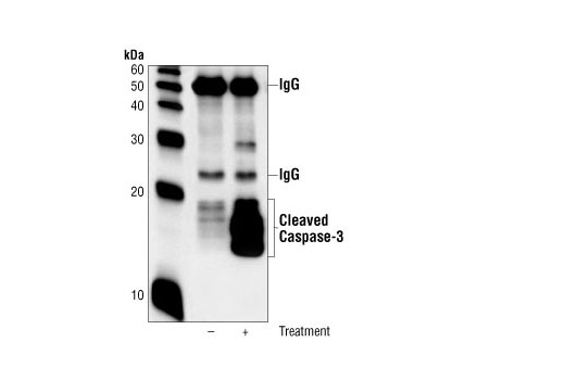
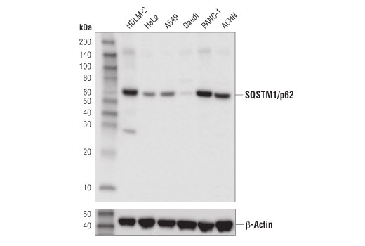
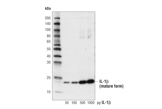
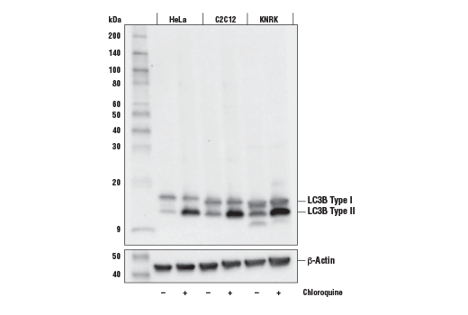
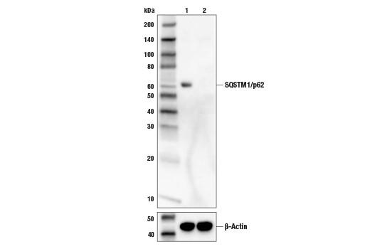
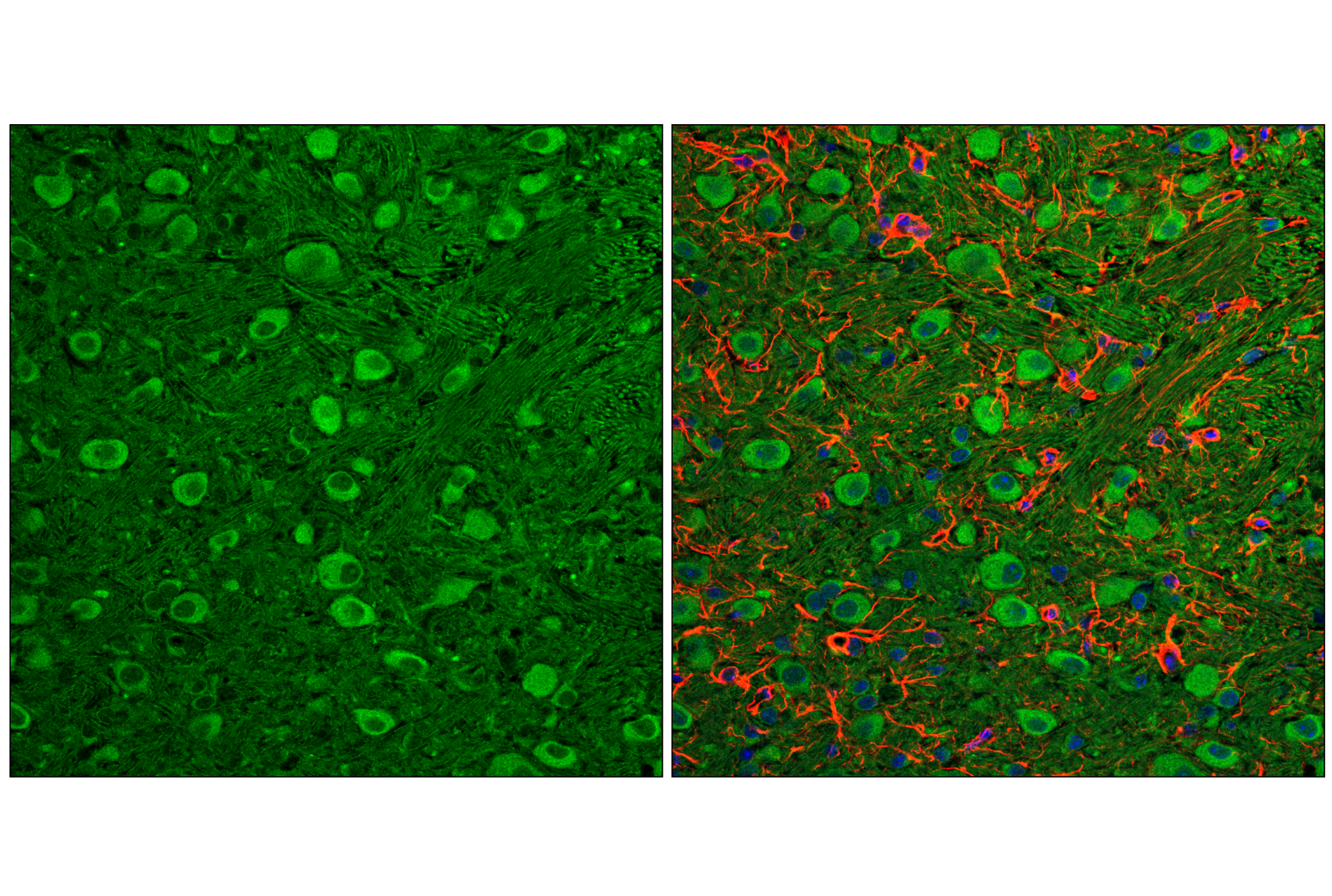
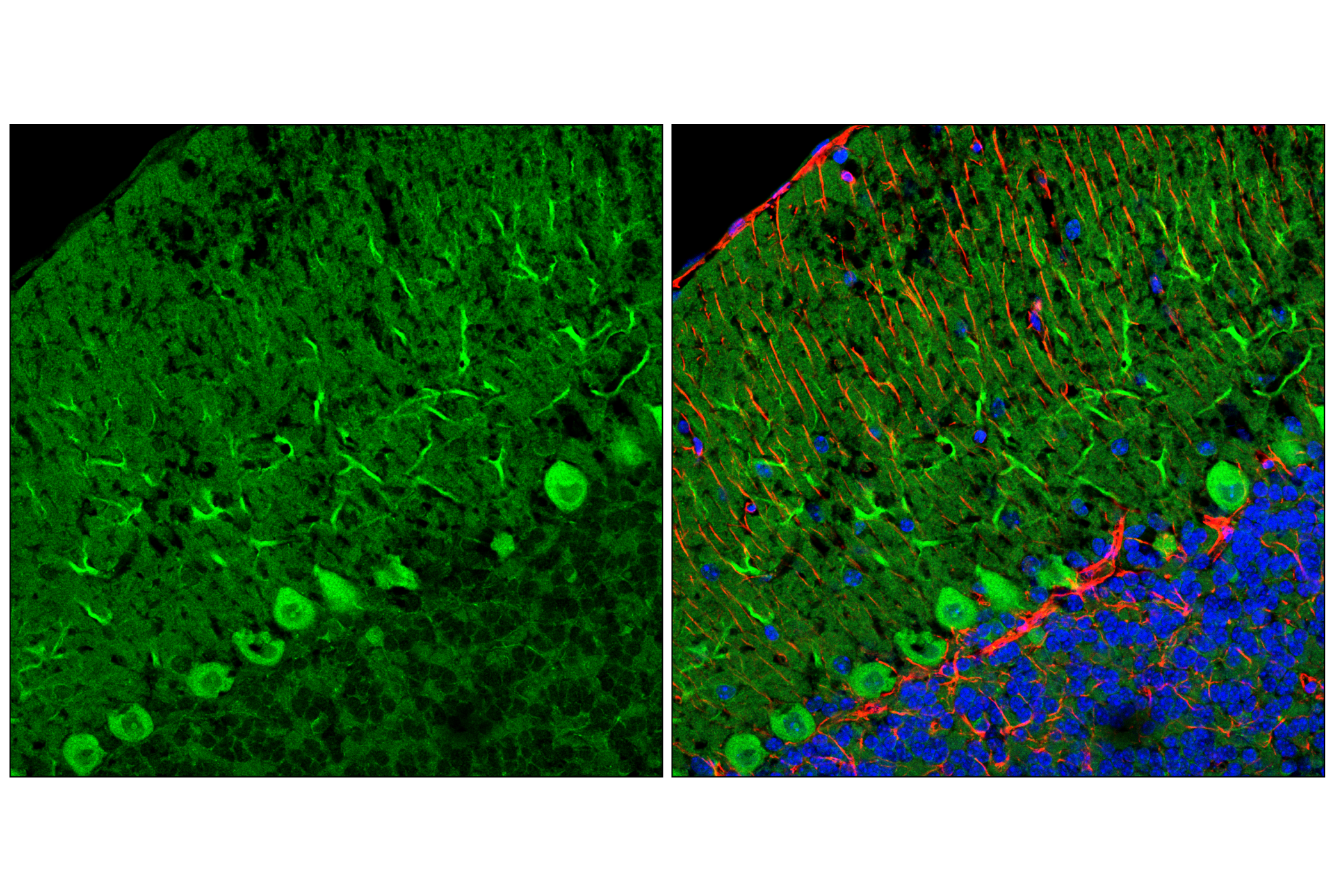
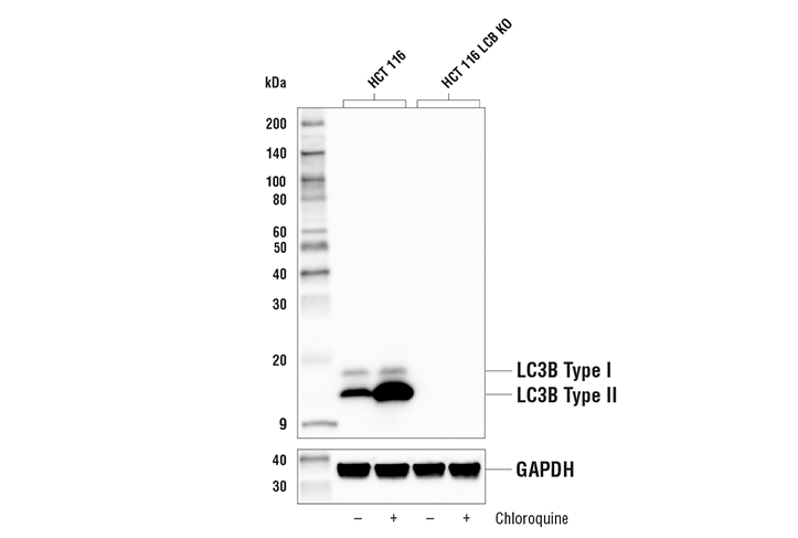
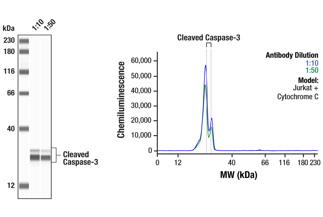
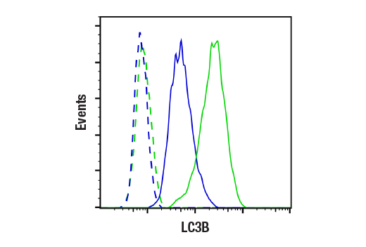
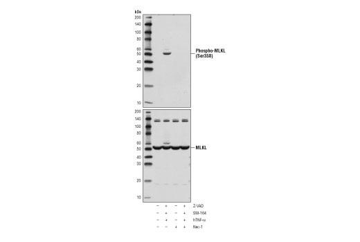
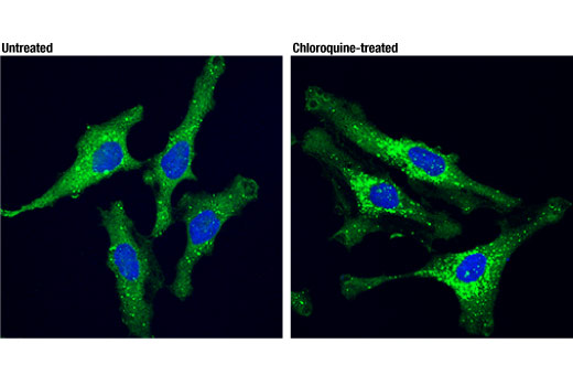
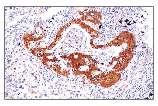
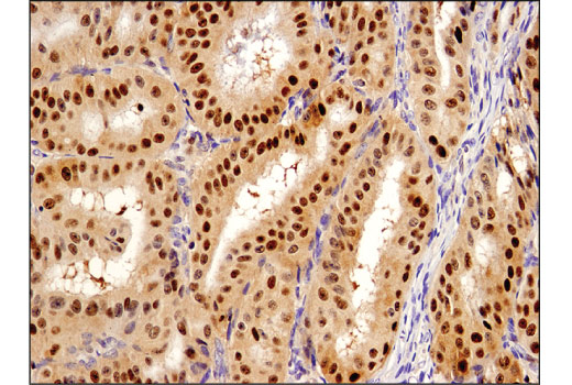
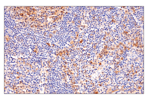
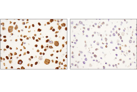
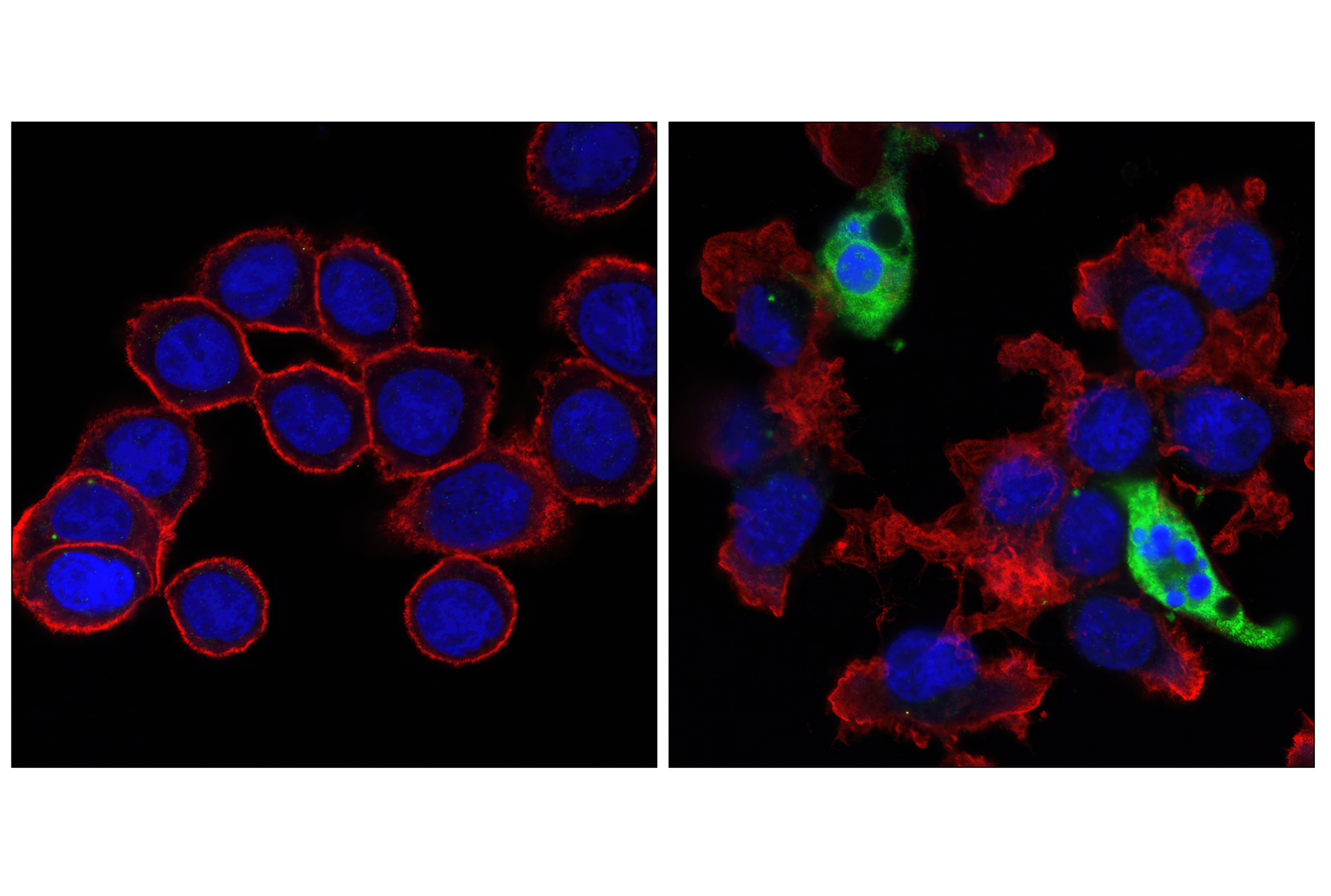
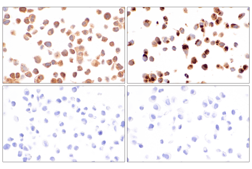
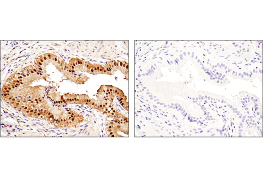
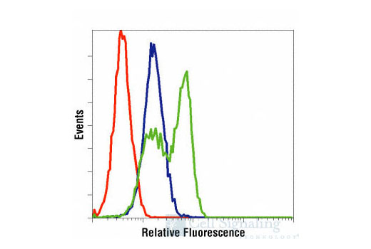
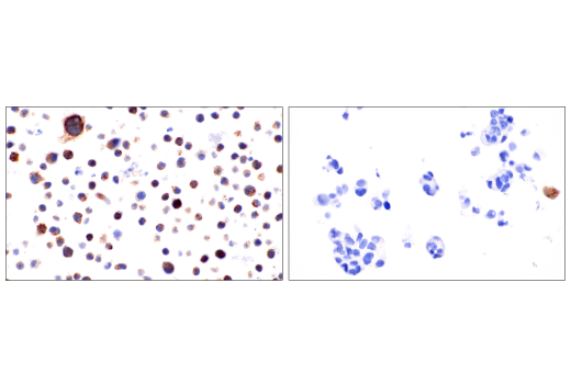
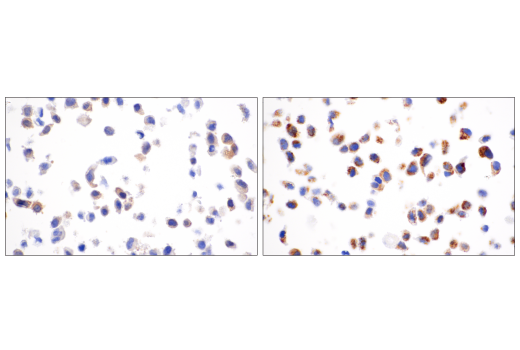
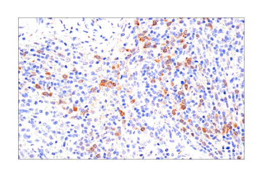
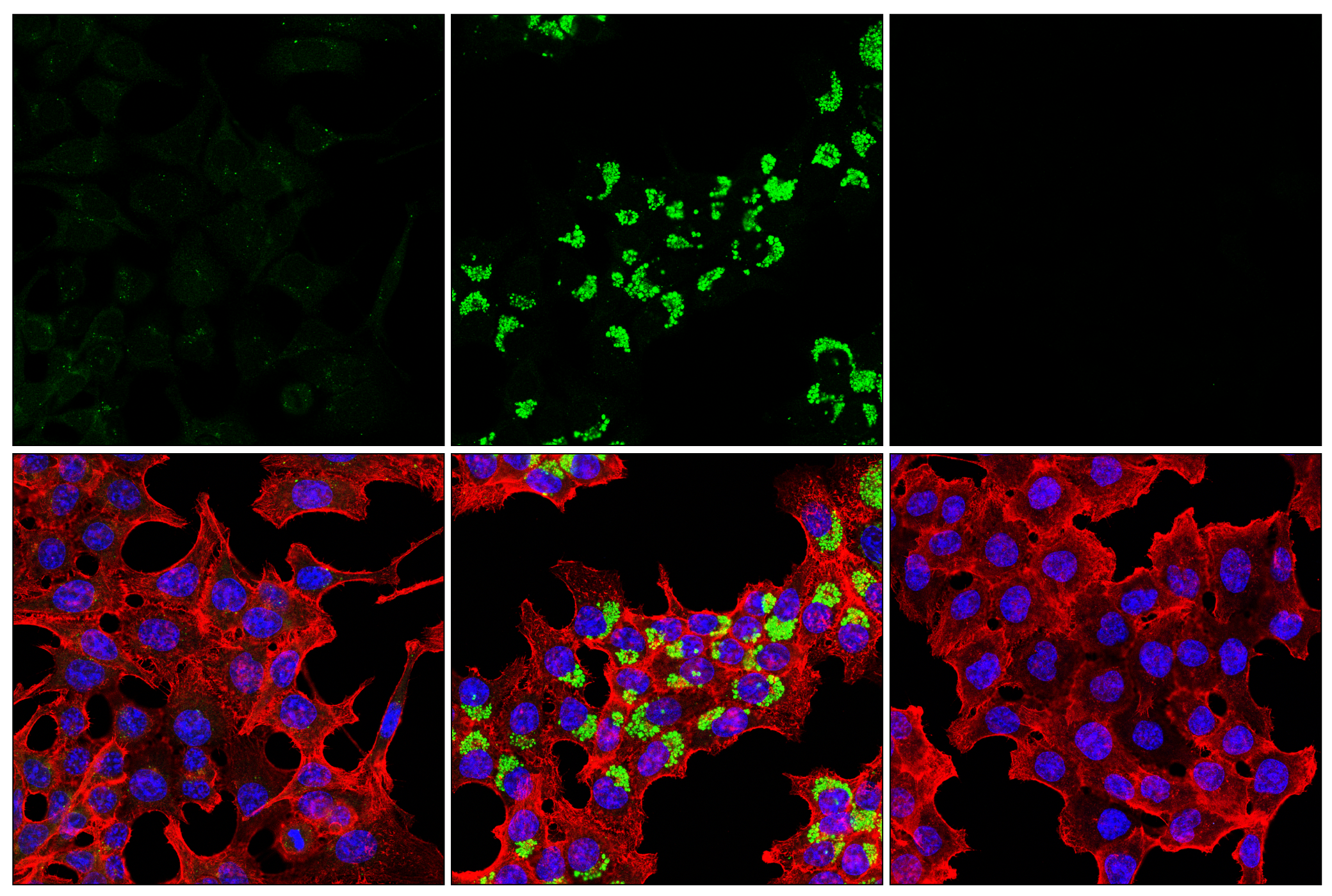
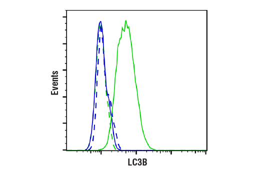
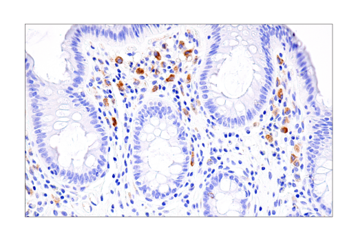
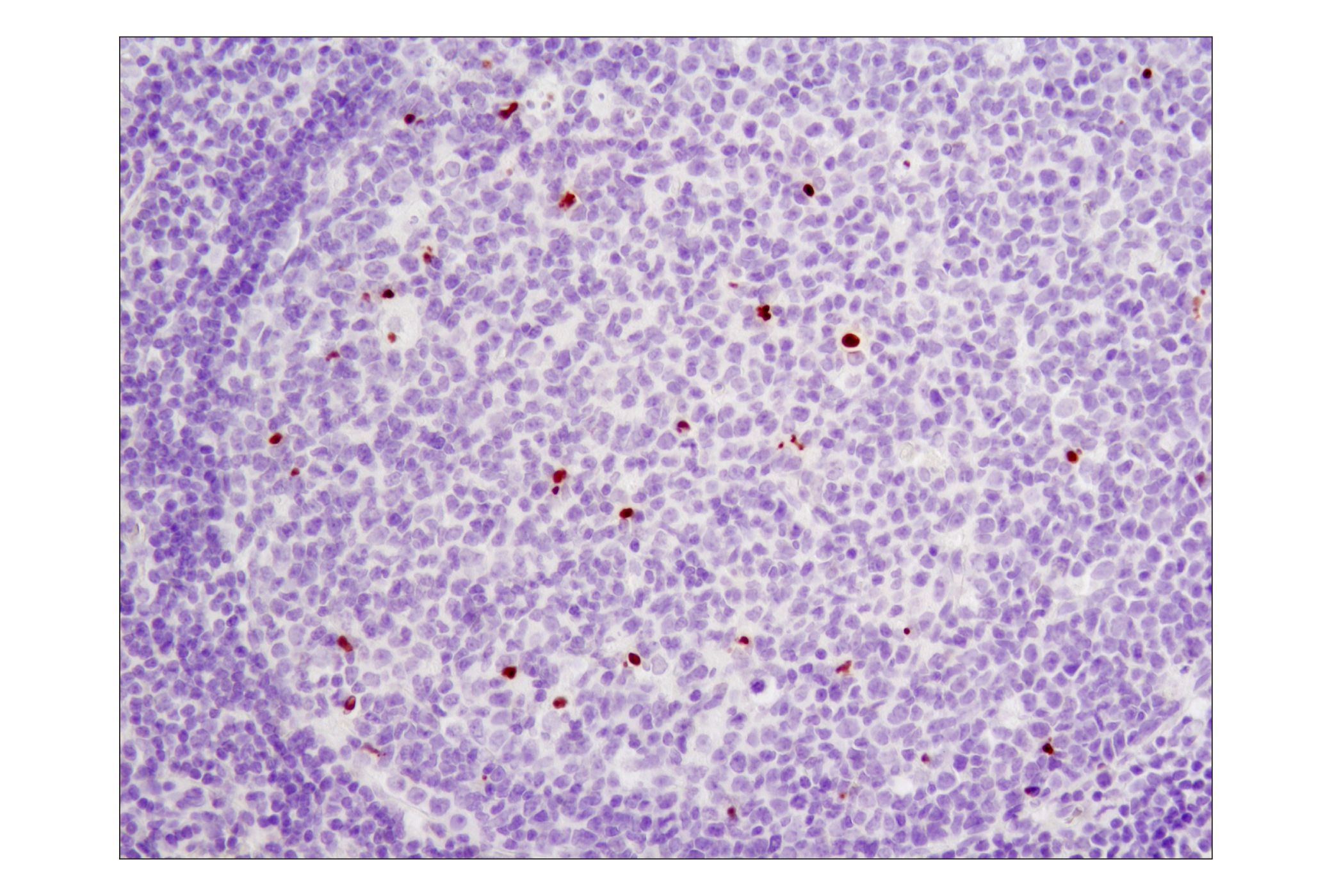
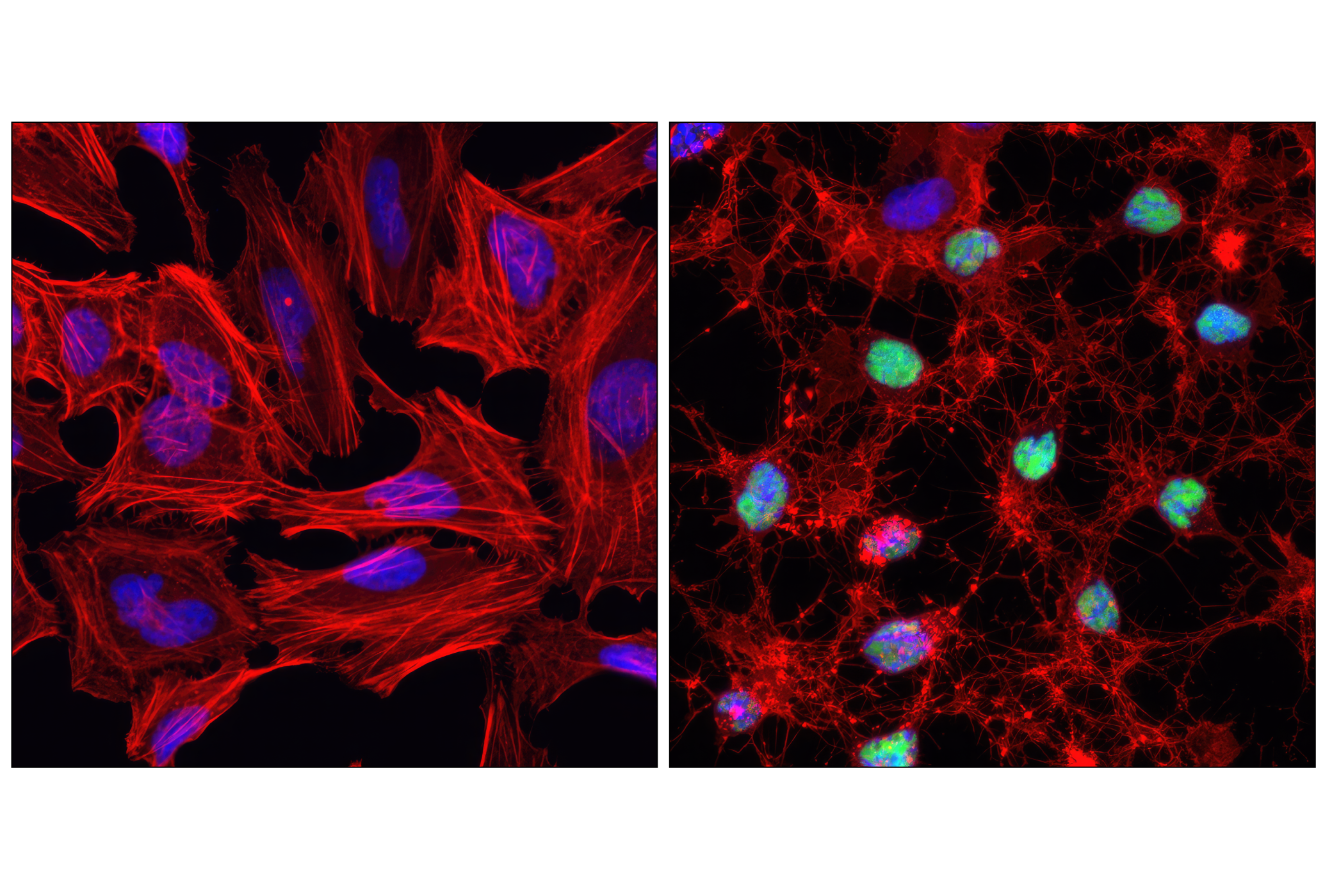
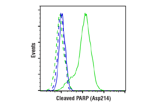
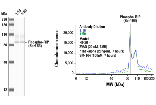
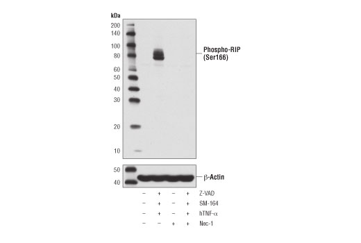
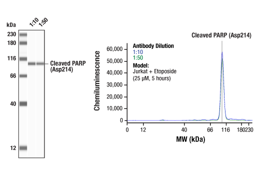
Each antibody in the Human Reactive Cell Death and Autophagy Antibody Sampler Kit detects endogenous levels of its target protein. Cleaved Caspase-3 (Asp175) (5A1E) Rabbit mAb detects endogenous levels of the large fragment (17/19 kDa) of activated caspase-3 resulting from cleavage adjacent to Asp175. This antibody does not recognize full-length caspase-3 or other cleaved caspases. Non-specific labeling may be observed by immunofluorescence in specific sub-types of healthy cells in fixed-frozen tissues (e.g., pancreatic alpha-cells). Cytoplasmic background may be observed in human and monkey samples. Cleaved PARP (Asp214) (D64E10) XP® Rabbit mAb detects endogenous levels of the large fragment (89 kDa) of human PARP1 protein produced by caspase cleavage. The antibody does not recognize full-length PARP1 or other PARP isoforms. Cleaved Gasdermin D (Asp275) (E7H9G) Rabbit mAb recognizes endogenous levels of the amino-terminal fragment of Gasdermin D only when cleaved at Asp275. Cleaved-IL-1β (Asp116) (D3A3Z) Rabbit mAb recognizes endogenous levels of mature IL-1β protein only when cleaved at Asp116. Phospho-RIP3 (Ser227) (D6W2T) Rabbit mAb detected a band at 30 kDa that appears to be a cleavage product of RIP3. Phospho-MLKL (Ser358) (D6H3V) Rabbit mAb may also bind to MLKL when dually phosphorylated at Thr357 and Ser358. LC3B (E5Q2K) Mouse mAb detects both type I and type II forms of LC3B. Cross reactivity was not detected with other family members.
背景
背景
Regulated cell death has been classified based on distinct morphological and biochemical pathways (1). Type I cell death, or apoptosis, is characterized by cytoplasmic shrinkage, chromatin condensation, nuclear fragmentation, plasma membrane blebbing, and phagocytic update of dead cells. Apoptosis can occur through extrinsic pathways involving extracellular factors, including the activation of death receptors, or through intrinsic pathways involving intracellular perturbations, including mitochondrial outer membrane permeabilization (2). Both of these apoptotic pathways lead to activation of caspases, a family of cysteine acid proteases that are synthesized as inactive zymogens containing pro-domains, followed by large (p20) and small (p10) subunits which are proteolytically activated in a cascade-like fashion. Caspase-3 is a key downstream protease activated by both extrinsic and intrinsic apoptotic pathways and cleaves a large number of proteins involved in the disassembly of the cell, including poly(ADP-ribose) polymerase (PARP), a protein involved in the DNA damage response. Type II cell death, or autophagy, manifests with extensive cytoplasmic vacuolization, and like apoptosis, can include phagocytic update. Autophagy is a catabolic process for the degradation of cellular components including protein aggregates, damaged organelles, and pathogens (3). The process involves the engulfment of these components into a double membrane structure, the autophagosome, which fuses to the lysosome for degradation. Autophagy requires, and can be monitored by, the conversion of LC3 family members, such as LC3B, from a type I form to a lipidated type II form that is incorporated into the autophagosome membrane and binds to a variety of cargo receptors. Cargo receptors such as SQSTM1/p62 bind LC3 along with ubiquitinated proteins that are targeted for degradation. SQSTM1/p62 is also degraded during this process, and thus its expression is frequently used to monitor this process.Type III cell death, or necrosis, manifests with plasma membrane permeability with cellular swelling and fragmentation, and lacks a clear phagocytic response which then leads to an inflammatory signaling with the release of damage-associated molecular patterns (DAMPs). Necrosis can be triggered by multiple regulated pathways including necroptosis and pyroptosis. Necroptosis is regulated by the kinase activities of RIP and RIP3 and the pore forming ability of MLKL (4). Necroptosis requires the activation of RIP3 which then phosphorylates MLKL at Ser358 (Ser345 in mouse). Phosphorylation of MLKL leads to generation of a pore complex involved in cell swelling and the secretion of DAMPs. RIP3 activation is triggered through several RIP homotypic interaction motif (RHIM) domain interactions including RIP, TRIF, and ZBP1 and results in the phosphorylation of RIP3 at Ser227 (Thr231/Ser232 in mouse). Canonical necroptosis signaling is mediated by RIP, and this can be inhibited by necrostatins, small molecules that directly inhibit RIP kinase activity. Activation of RIP can be monitored through autophosphorylation sites including Ser166. Pyroptosis is generally induced in cells of the innate immune system, and is characterized by cleavage of Gasdermin D (5). The amino-terminal fragment of Gasdermin D produced following cleavage by inflammatory caspases (Caspase-1, -4, -5), oligomerizes to form a pore. Canonical cleavage of Gasdermin D occurs through a two-step process. The first step involves transcriptional regulation of targets such as NLRP3 and the pro-forms of IL-1β and IL-18. In the second execution step, Caspase-1 is activated through formation of inflammasome complexes. Activated Caspase-1 cleaves Gasdermin D as well as IL-1β and IL-18 to their mature forms, and these active cytokines are secreted through pores formed by Gasdermin D.
1.Galluzzi, L. et al. (2018) Cell Death Differ 25, 486-541.
2.Green, D.R. (1998) Cell 94, 695-8.
3.Codogno, P. and Meijer, A.J. (2005) Cell Death Differ 12 Suppl 2, 1509-18.
4.Shan, B. et al. (2018) Genes Dev 32, 327-40.
5.Shi, J. et al. (2017) Trends Biochem Sci 42, 245-54.
研究领域
癌症,细胞生物学,纤维化,免疫学和肿瘤学,代谢,神经科学
数据库链接
Entrez-Gene ID
8737,3553,8878,11035,79792,197259,836,81631,142
UniProt ID
Q13546,P01584,Q13501,Q9Y572,P57764,Q8NB16,P42574,Q9GZQ8,P09874
声明 :本官网所有报价均为常温或者蓝冰运输价格,如有产品需要干冰运输,需另外加收干冰运输费。

 危险品化学品经营许可证(不带存储) 许可证编号:沪(杨)应急管危经许[2022]202944(QY)
  营业执照(三证合一)