


 全部商品分类
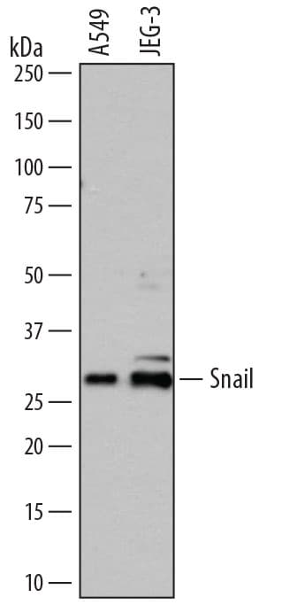


 下载产品说明书 下载SDS
 用小程序,查商品更便捷


 收藏
收藏
 对比
对比 咨询
咨询Chromatin Immunoprecipitation (ChIP)(5 µg/5 x 106 cells)
Immunocytochemistry(5-15 µg/mL)

Pro2-Arg264
Accession # O95863
Scientific Data
 View Larger
Detection of Human Snail by Western Blot. Western blot shows lysates of A549 human lung carcinoma cell line and JEG-3 human epithelial choriocarcinoma cell line. PVDF membrane was probed with 0.5 µg/mL of Goat Anti-Human Snail Antigen Affinity-purified Polyclonal Antibody (Catalog # AF3639) followed by HRP-conjugated Anti-Goat IgG Secondary Antibody (Catalog # HAF109). A specific band was detected for Snail at approximately 29 kDa (as indicated). This experiment was conducted under reducing conditions and using Immunoblot Buffer Group 1.
 View Larger
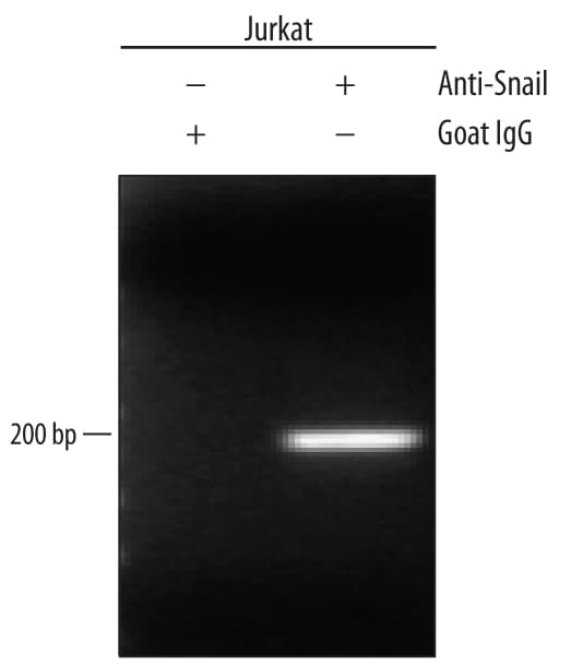
Detection of Snail-regulated Genes by Chromatin Immunoprecipitation. Jurkat human acute T cell leukemia cell line treated with 50 ng/mL PMA and 200 ng/mL calcium ionomycin for 30 minutes was fixed using formaldehyde, resuspended in lysis buffer, and sonicated to shear chromatin. Snail/DNA complexes were immunoprecipitated using 5 µg Goat Anti-Human Snail Antigen Affinity-purified Polyclonal Antibody (Catalog # AF3639) or control antibody (Catalog # AB-108-C) for 15 minutes in an ultrasonic bath, followed by Biotinylated Anti-Goat IgG Secondary Antibody (Catalog # BAF109). Immunocomplexes were captured using 50 µL of MagCellect Streptavidin Ferrofluid (Catalog # MAG999) and DNA was purified using chelating resin solution. TheE-Cadherinpromoter was detected by standard PCR.
.") View Larger
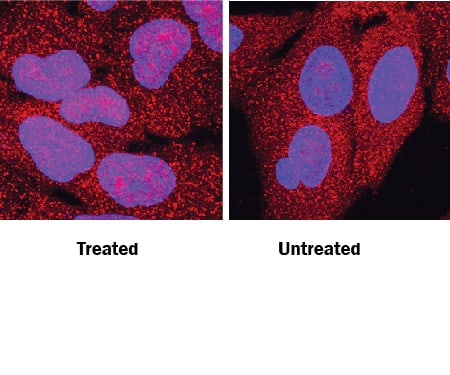
View LargerSnail in A549 Human Cell Line. Snail was detected in immersion fixed A549 human lung carcinoma cell line treated with Recombinant Human TGF-beta 1 (left panel, Catalog # 240-B) or untreated (right panel) using Goat Anti-Human Snail Antigen Affinity-purified Polyclonal Antibody (Catalog # AF3639) at 10 µg/mL for 3 hours at room temperature. Cells were stained using the NorthernLights™ 557-conjugated Anti-Goat IgG Secondary Antibody (red; Catalog # NL001) and counterstained with DAPI (blue). Specific staining was localized to nuclei. View our protocol for Fluorescent ICC Staining of Cells on Coverslips.
 View Larger
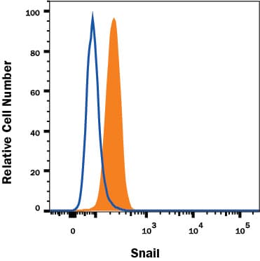
Detection of Snail in A549 Human Cell Line by Flow Cytometry. A549 human lung carcinoma cell line was stained with Goat Anti-Human Snail Antigen Affinity-purified Polyclonal Antibody (Catalog # AF3639, filled histogram) or isotype control antibody (Catalog # AB-108-C, open histogram), followed by Fluorescein-conjugated Anti-Goat IgG Secondary Antibody (Catalog # F0109). To facilitate intracellular staining, cells were fixed and permeabilized with FlowX FoxP3 Fixation & Permeabilization Buffer Kit (Catalog # FC012). View our protocol for Staining Intracellular Molecules.
 View Larger
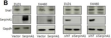
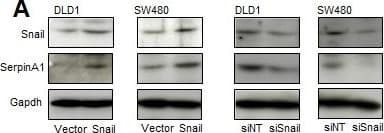
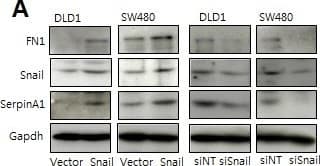
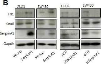
Detection of Human Snail by Western Blot SerpinA1 was regulated by SnailA. DLD-1 and SW480 cells were transfected with pcDNA-Snail (Snail), control vector pcDNA (vector), Snail siRNA (siSnail), or nontargeting siRNA (siNT), and Snail and serpinA1 protein levels were evaluated by western blot analysis. B. DLD-1 and SW480 cells were transfected with pcDNA-serpinA1 (serpinA1), control vector pcDNA (vector), serpinA1 siRNA (siSerpinA1), or nontargeting siRNA (siNT), and western blot analysis was performed for detection of Snail and SerpinA1 expression. C. DLD-1 and SW480 cells were transfected with pcDNA-Snail (Snail) or control vector pcDNA (vector), and ChIP assays were performed. The presence of the serpinA1 promoter (−516/−4) was verified in immunoprecipitates with either mouse IgG or anti-Snail antibodies, and assay inputs were analyzed using real-time PCR. The samples were loaded on agarose gels. D. Data show promoter enrichment in the anti-Snail immunoprecipitate relative to IgG. Image collected and cropped by CiteAb from the following publication (https://pubmed.ncbi.nlm.nih.gov/26015410), licensed under a CC-BY license. Not internally tested by R&D Systems.
 View Larger
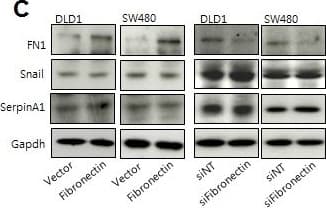
Detection of Human Snail by Western Blot Snail and serpinA1 promoted tumor progression through fibronectinA. DLD-1 and SW480 cells were transfected with pcDNA-Snail (Snail), control vector pcDNA (vector), Snail siRNA (siSnail), or nontargeting siRNA (siNT), and Snail, serpinA1, and fibronectin protein levels were evaluated by western blot analysis. B. DLD-1 and SW480 cells were transfected with pcDNA-serpinA1 (SerpinA1), control vector pcDNA (vector), serpinA1 siRNA (siSerpinA1), or nontargeting siRNA (siNT), and Snail, serpinA1, and fibronectin protein levels were evaluated by western blot analysis. C. DLD-1 and SW480 cells were transfected with pcDNA-fibronectin (Fibronectin), control vector pcDNA (vector), fibronectin siRNA (siFibronectin), or nontargeting siRNA (siNT), and Snail, serpinA1, and fibronectin protein levels were evaluated by western blot analysis. D., E. Invasion and migration assays were performed using transfected cells. Representative data are shown for cells that invaded (top) and migrated (bottom) in the presence of 1% FBS. *P < 0.05. Image collected and cropped by CiteAb from the following publication (https://pubmed.ncbi.nlm.nih.gov/26015410), licensed under a CC-BY license. Not internally tested by R&D Systems.
 View Larger
Detection of Human Snail by Western Blot Snail and serpinA1 promoted tumor progression through fibronectinA. DLD-1 and SW480 cells were transfected with pcDNA-Snail (Snail), control vector pcDNA (vector), Snail siRNA (siSnail), or nontargeting siRNA (siNT), and Snail, serpinA1, and fibronectin protein levels were evaluated by western blot analysis. B. DLD-1 and SW480 cells were transfected with pcDNA-serpinA1 (SerpinA1), control vector pcDNA (vector), serpinA1 siRNA (siSerpinA1), or nontargeting siRNA (siNT), and Snail, serpinA1, and fibronectin protein levels were evaluated by western blot analysis. C. DLD-1 and SW480 cells were transfected with pcDNA-fibronectin (Fibronectin), control vector pcDNA (vector), fibronectin siRNA (siFibronectin), or nontargeting siRNA (siNT), and Snail, serpinA1, and fibronectin protein levels were evaluated by western blot analysis. D., E. Invasion and migration assays were performed using transfected cells. Representative data are shown for cells that invaded (top) and migrated (bottom) in the presence of 1% FBS. *P < 0.05. Image collected and cropped by CiteAb from the following publication (https://pubmed.ncbi.nlm.nih.gov/26015410), licensed under a CC-BY license. Not internally tested by R&D Systems.
Human Snail Antibody Summary
Pro2-Arg264
Accession # O95863
Applications
Please Note: Optimal dilutions should be determined by each laboratory for each application. General Protocols are available in the Technical Information section on our website.
Chromatin Immunoprecipitation (ChIP)(5 µg/5 x 106 cells)
Immunocytochemistry(5-15 µg/mL)
Background: Snail
Snail is predicted 29 kDa nuclear zinc finger transcriptional repressor that contains an N-terminal basic SNAG domain followed by three classical and one atypical zinc finger domains. During development, Snail is required for the establishment of left-right axis asymmetry. It also regulates the transcription of E-cadherin and other genes involved in epithelial-mesenchymal transitions during cancer progression. Human Snail shares 88% amino acid sequence identity with mouse and rat Snail.
Preparation and Storage
- 12 months from date of receipt, -20 to -70 °C as supplied.
- 1 month, 2 to 8 °C under sterile conditions after reconstitution.
- 6 months, -20 to -70 °C under sterile conditions after reconstitution.
参考图片
Detection of Human Snail by Western Blot. Western blot shows lysates of A549 human lung carcinoma cell line and JEG‑3 human epithelial choriocarcinoma cell line. PVDF membrane was probed with 0.5 µg/mL of Goat Anti-Human Snail Antigen Affinity-purified Polyclonal Antibody (Catalog # AF3639) followed by HRP-conjugated Anti-Goat IgG Secondary Antibody (Catalog # HAF109). A specific band was detected for Snail at approximately 29 kDa (as indicated). This experiment was conducted under reducing conditions and using Immunoblot Buffer Group 1.
Detection of Snail-regulated Genes by Chromatin Immunoprecipitation. Jurkat human acute T cell leukemia cell line treated with 50 ng/mL PMA and 200 ng/mL calcium ionomycin for 30 minutes was fixed using formaldehyde, resuspended in lysis buffer, and sonicated to shear chromatin. Snail/DNA complexes were immunoprecipitated using 5 μg Goat Anti-Human Snail Antigen Affinity-purified Polyclonal Antibody (Catalog # AF3639) or control antibody (Catalog # AB-108-C) for 15 minutes in an ultrasonic bath, followed by Biotinylated Anti-Goat IgG Secondary Antibody (Catalog # BAF109). Immunocomplexes were captured using 50 μL of MagCellect Streptavidin Ferrofluid (Catalog # MAG999) and DNA was purified using chelating resin solution. The E-Cadherin promoter was detected by standard PCR.
Snail in A549 Human Cell Line. Snail was detected in immersion fixed A549 human lung carcinoma cell line treated with Recombinant Human TGF-beta 1 (left panel, Catalog # 240-B) or untreated (right panel) using Goat Anti-Human Snail Antigen Affinity-purified Polyclonal Antibody (Catalog # AF3639) at 10 µg/mL for 3 hours at room temperature. Cells were stained using the NorthernLights™ 557-conjugated Anti-Goat IgG Secondary Antibody (red; Catalog # NL001) and counterstained with DAPI (blue). Specific staining was localized to nuclei. View our protocol for Fluorescent ICC Staining of Cells on Coverslips.