


 全部商品分类
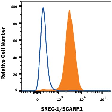


hSREC-I MAb (Cl 3736 (25 ug)

 下载产品说明书 下载SDS
 用小程序,查商品更便捷


 收藏
收藏
 对比
对比 咨询
咨询Flow Cytometry(0.25 µg/106 cells)

Ser20-Thr421
Accession # Q14162
Scientific Data
 View Larger
Detection of SREC‑I/SCARF1 in HUVEC Human Cells by Flow Cytometry. HUVEC human umbilical vein endothelial cells were stained with Mouse Anti-Human SREC-I/SCARF1 Monoclonal Antibody (Catalog # MAB2409, filled histogram) or isotype control antibody (Catalog # MAB0041, open histogram), followed by Phycoerythrin-conjugated Anti-Mouse IgG Secondary Antibody (Catalog # F0102B). View our protocol for Staining Membrane-associated Proteins.
Human SREC-I/SCARF1 Antibody Summary
Ser20-Thr421
Accession # Q14162
Applications
Please Note: Optimal dilutions should be determined by each laboratory for each application. General Protocols are available in the Technical Information section on our website.
Flow Cytometry(0.25 µg/106 cells)
Background: SREC-I/SCARF1
The scavenger receptor (SR) family comprises a group of functionally defined membrane receptors that share a common ability to bind and internalize modified forms of low density lipoproteins (LDL) such as acetylated LDL (AcLDL) and oxidized LDL(OxLDL) (1‑3). Family members are classified alphabetically. They play important roles in lipid metabolism, in host defence and in the regulation of acquired immunity (2, 4). Scavenger receptor expressed by endothelial cells-I (SREC-I; also called SCARF1) and SREC-2 are two proteins that belong to the F type scavenger receptor group (SR-F1 and SR-F2). The full length cDNA of human SREC-I encodes an 830 amino acid (aa) type I transmembrane protein which contains a 19 aa signal peptide, a 402 aa extracellular region, a 21 aa transmembrane segment, and a 388 aa long cytoplasmic domain. The extracellular region contains ten EGF-like repeats (five of which fit the exact consensus sequence for an EGF-like domain) while the cytoplasmic domain is rich in serine and proline in the N-terminal half, and glycine in the C-terminal segment (5, 6). In addition to the full length form, four SREC-I isoforms exist. Two show insertions of a stop codon in EGF-like domain 8, resulting in mature soluble forms of 323 aa and 318 aa, respectively. A third isoform deletes part of domain 8 plus domains 9 and 10; it continues in-frame to generate a mature transmembrane protein of 725 aa. The last isoform shows only cytoplasmic splicing, with 72 aa substituted for the last 332 aa of the full length form. All three transmembrane forms bind acetylated LDL (6). Native SREC-I is approximately 150 kDa and is expressed by endothelial cells, macrophages and fetal neurons (7, 8). In the extracellular region, human SREC-I shares 76% and 53% aa sequence identity with mouse SREC-I and human SREC-2, respectively.
- Horiuchi, S. et al. (2003) Amino Acids 25:283.
- Greaves, D.R. and S. Gordon (2005) J. Lipid Res. 46:11.
- Platt, N. and S. Gordon (1998) Chem. Biol. 5:R193.
- Platt, N. and S. Gordon (2001) J. Clin. Invest. 108:649.
- Adachi, H. et al. (1997) J. Biol. Chem. 272:31217.
- Adachi, H. and M. Tsujimoto (2002) J. Biol. Chem. 277:24014.
- Shibata, M. et al. (2004) J. Biol. Chem. 279:40084.
- Tanura, Y. et al. (2004) J. Biol. Chem. 279:30938.
Preparation and Storage
- 12 months from date of receipt, -20 to -70 °C as supplied.
- 1 month, 2 to 8 °C under sterile conditions after reconstitution.
- 6 months, -20 to -70 °C under sterile conditions after reconstitution.
参考图片
Detection of SREC‑I/SCARF1 in HUVEC Human Cells by Flow Cytometry. HUVEC human umbilical vein endothelial cells were stained with Mouse Anti-Human SREC‑I/SCARF1 Monoclonal Antibody (Catalog # MAB2409, filled histogram) or isotype control antibody (Catalog # MAB0041, open histogram), followed by Phycoerythrin-conjugated Anti-Mouse IgG Secondary Antibody (Catalog # F0102B). View our protocol for Staining Membrane-associated Proteins.