


 全部商品分类
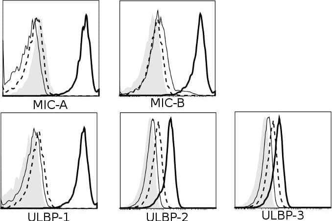


 下载产品说明书 下载SDS
 用小程序,查商品更便捷


 收藏
收藏
 对比
对比 咨询
咨询
Scientific Data
 View Larger
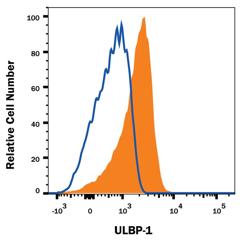
Detection of ULBP‑1 in MOLT‑4 Human Cell Line by Flow Cytometry. MOLT-4 human acute lymphoblastic leukemia cell line was stained with Mouse Anti-Human ULBP-1 Monoclonal Antibody (Catalog # MAB1380, filled histogram) or isotype control antibody (Catalog # MAB003, open histogram), followed by Phycoerythrin-conjugated Anti-Mouse IgG Secondary Antibody (Catalog # F0102B).
 View Larger
Detection of Human ULBP-1 by Flow Cytometry Multiple receptors and ligands are involved in NK cell-mediated lysis of activated CD4+ T cells.Role of (A) activating and (B) inhibitory NK receptors in NK cell degranulation. Left column: representative histograms (of n≥3) for surface expression of ligands on activated (thick black line) and resting CD4+ T cells (thin black line). Isotype-matched control Ig are represented by dashed line (activated CD4+ T) and filled histogram (resting CD4+ T). Middle- and right column: NK and CD4+ T cells were activated for 4 days in vitro as described, and co-cultured for 4 hours with 10 ug/mL mAb (or relevant isotype-matched control Ig). Degranulation is shown for CD56dim (middle column) and CD56bright (right column) NK cells. Representative histograms of surface expression of receptors on activated (thick black line) and resting NK cells (thin black line). Isotype-matched control Ig are represented by dashed line (activated NK) and filled histogram (resting NK). * P<0.05, ** P<0.005, *** P<0.001. (C) Sorted IL-2-activated CD56dim and CD56bright NK cells were co-cultured with 51Cr-labeled activated CD4+ T cells in a 51Cr-release assay with human IgG4 isotype control (•) or anti-NKG2A mAb (○). Data represents n = 3 experiments. Image collected and cropped by CiteAb from the following publication (https://pubmed.ncbi.nlm.nih.gov/22384114), licensed under a CC-BY license. Not internally tested by R&D Systems.
Human ULBP-1 Antibody Summary
Applications
Please Note: Optimal dilutions should be determined by each laboratory for each application. General Protocols are available in the Technical Information section on our website.
Background: ULBP-1
ULBP-1 is a member of a family of cell-surface proteins that function as ligands for human NKG2D. ULBP-1 has also been described under the names RaeT1I (retinoic acid early transcript), ALCAN-beta, and NKG2DL1. The name ULBP-1 derives from the original identification of three proteins, ULBP-1, -2, and -3, as ligands for the human cytomegalovirus glycoprotein UL16; they were designated UL16 binding proteins (ULBP). The gene for ULBP-1 resides in a cluster of ten related genes, six of which encode potentially functional glycoproteins. Amino acid sequence identity within this family ranges from 30-95%. These proteins are distantly related to MHC class I proteins, but they possess only the alpha 1 and alpha 2 Ig-like domains, and they have no capacity to bind peptide or interact with beta 2-microglobulin. They are anchored to the membrane via a GPI-linkage. ULBP-1 and several other family members are known to bind to human NKG2D, an activating receptor expressed on NK cells, NKT cells, gamma δ T cells, and CD8+ alpha beta T cells. Engagement of NKG2D results in the activation of cytolytic activity and/or cytokine production by these effector cells. ULBP-1 is expressed on some tumor cells and has been implicated in tumor surveillance (1-8).
- Cosman, D. et al. (2001) Immunity 14:123.
- Kubin, M. et al. (2001) Eur. J. Immunol. 31:1428.
- Sutherland, C. et al. (2002) J. Immunol. 168:671.
- Steinle, A. et al. (2001) Immunogenetics 53:279.
- Sutherland, C. et al. (2001) Immunol. Rev. 181:185
- Pende, D. et al. (2002) Cancer Res. 62:6178.
- Radosavljevic, M. et al. (2002) Genomics 79:114
- NKG2D and its Ligands, 2002; www.RnDSystems.com.
Preparation and Storage
- 12 months from date of receipt, -20 to -70 °C as supplied.
- 1 month, 2 to 8 °C under sterile conditions after reconstitution.
- 6 months, -20 to -70 °C under sterile conditions after reconstitution.
参考图片
Detection of ULBP‑1 in MOLT‑4 Human Cell Line by Flow Cytometry. MOLT‑4 human acute lymphoblastic leukemia cell line was stained with Mouse Anti-Human ULBP‑1 Monoclonal Antibody (Catalog # MAB1380, filled histogram) or isotype control antibody (Catalog # MAB003, open histogram), followed by Phycoerythrin-conjugated Anti-Mouse IgG Secondary Antibody (Catalog # F0102B).