


 全部商品分类
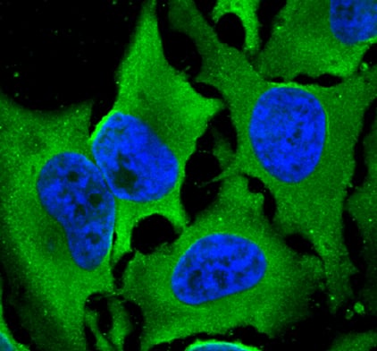


 下载产品说明书 下载SDS
 用小程序,查商品更便捷


 收藏
收藏
 对比
对比 咨询
咨询
Scientific Data
.") View Larger
View LargerLaminin S in HeLa Human Cell Line. Laminin S was detected in immersion fixed HeLa human cervical epithelial carcinoma cell line using Mouse Anti-Human/Rat/Chicken Laminin S Monoclonal Antibody (Catalog # MAB2066) at 10 µg/mL for 3 hours at room temperature. Cells were stained using the NorthernLights™ 493-conjugated Anti-Mouse IgG Secondary Antibody (green; Catalog # NL009) and counterstained with DAPI (blue). Specific staining was localized to cytoplasm. View our protocol for Fluorescent ICC Staining of Cells on Coverslips.
Human/Rat/Chicken Laminin S Antibody Summary
Applications
Green, T.L. et al. (1992) J. Biol. Chem. 267:2014. This application was not tested by R&D Systems.
Please Note: Optimal dilutions should be determined by each laboratory for each application. General Protocols are available in the Technical Information section on our website.
Background: Laminin S
Laminins are heterotrimeric, noncollagenous glycoproteins composed of alpha, beta and gamma chains. Through interactions with integrins, dystroglycan and other receptors, laminins critically contribute to cell differentation, cell shape and migration and maintenance of tissue phenotypes and survival (1). Laminin S, also known as laminin beta 2, is a subunit of laminin-3 (S-laminin), laminin-4 (S-merosin) and laminin-7 (KS-laminin). It is enriched in the basement membrane of muscles at the neuromuscular junctions, kidney glomerulus and vascular smooth muscle.
Preparation and Storage
- 12 months from date of receipt, -20 to -70 °C as supplied.
- 1 month, 2 to 8 °C under sterile conditions after reconstitution.
- 6 months, -20 to -70 °C under sterile conditions after reconstitution.
参考图片
Laminin S in HeLa Human Cell Line. Laminin S was detected in immersion fixed HeLa human cervical epithelial carcinoma cell line using Mouse Anti-Human/Rat/Chicken Laminin S Monoclonal Antibody (Catalog # MAB2066) at 10 µg/mL for 3 hours at room temperature. Cells were stained using the NorthernLights™ 493-conjugated Anti-Mouse IgG Secondary Antibody (green; Catalog # NL009) and counterstained with DAPI (blue). Specific staining was localized to cytoplasm. View our protocol for Fluorescent ICC Staining of Cells on Coverslips.