


 全部商品分类


Iba1/AIF-1 (E4O4W) XP ® Rabbit mAb

 下载产品说明书 下载COA 下载SDS
 用小程序,查商品更便捷


 收藏
收藏
 对比
对比 咨询
咨询
Monoclonal antibody is produced by immunizing animals with a synthetic peptide corresponding to residues surrounding Ala139 of human Iba1/AIF-1 protein.
Product Usage Information
| Application | Dilution |
|---|---|
| Western Blotting | 1:1000 |
| Simple Western™ | 1:10 - 1:50 |
| Immunoprecipitation | 1:50 |
| IHC Leica Bond | 1:800 - 1:3200 |
| Immunohistochemistry (Paraffin) | 1:800 - 1:3200 |
| Immunofluorescence (Frozen) | 1:50 - 1:200 |
| Immunofluorescence (Immunocytochemistry) | 1:50 - 1:200 |
| Flow Cytometry (Fixed/Permeabilized) | 1:50 - 1:200 |
Specificity/Sensitivity
Species Reactivity:
Human, Mouse, Rat, Hamster, Monkey
Supplied in 10 mM sodium HEPES (pH 7.5), 150 mM NaCl, 100 µg/ml BSA, 50% glycerol and less than 0.02% sodium azide. Store at –20°C. Do not aliquot the antibody.
For a carrier-free (BSA and azide free) version of this product see product #79394.
参考图片
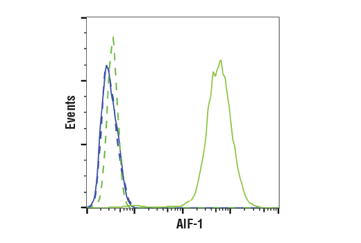
Flow cytometric analysis of SH-SY5Y cells (blue, negative) and THP-1 cells (green, positive) using Iba1/AIF-1 (E4O4W) XP® Rabbit mAb (solid lines) or a concentration-matched Rabbit (DA1E) mAb IgG XP® Isotype Control #3900 (dashed lines). Anti-rabbit IgG (H+L), F(ab')2 Fragment (Alexa Fluor® 488 Conjugate) #4412 was used as a secondary antibody.
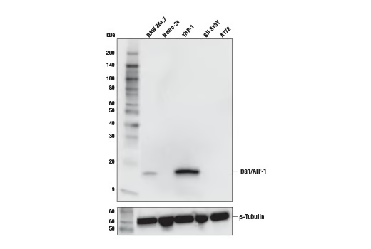
Western blot analysis of extracts from various cell lines using Iba1/AIF-1 (E4O4W) XP® Rabbit mAb (upper) and β-Tubulin (D2N5G) Rabbit mAb #15115 (lower).
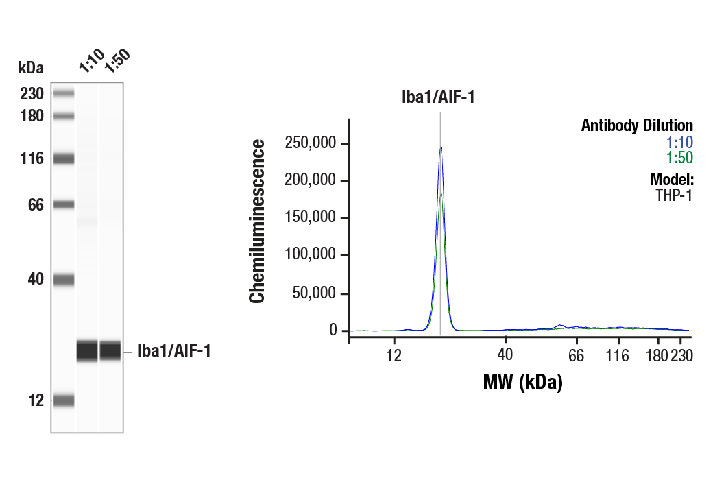
Simple Western™ analysis of lysates (1 mg/mL) from THP-1 cells using Iba1/AIF-1 (E4O4W) XP® Rabbit mAb #17198. The virtual lane view (left) shows the target band (as indicated) at 1:10 and 1:50 dilutions of primary antibody. The corresponding electropherogram view (right) plots chemiluminescence by molecular weight along the capillary at 1:10 (blue line) and 1:50 (green line) dilutions of primary antibody. This experiment was performed under reducing conditions on the Jess™ Simple Western instrument from ProteinSimple, a BioTechne brand, using the 12-230 kDa separation module.
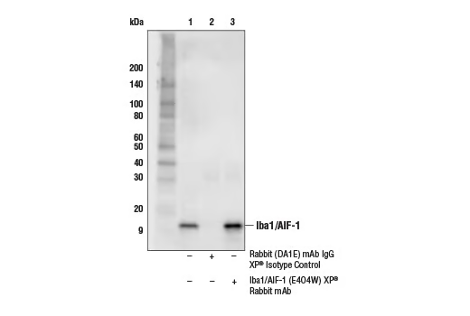
Immunoprecipitation of Iba1/AIF-1 from THP-1 cell extracts. Lane 1 is 10% input, lane 2 is Rabbit (DA1E) mAb IgG XP® Isotype Control #3900, and lane 3 is Iba1/AIF-1 (E4O4W) XP® Rabbit mAb. Western blot analysis was performed using Iba1/AIF-1 (E4O4W) XP® Rabbit mAb.
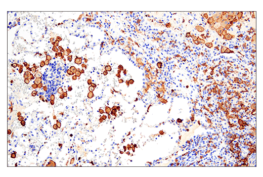
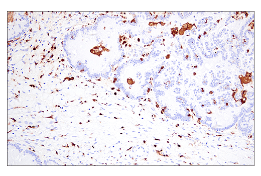
Immunohistochemical analysis of paraffin-embedded human neuroendocrine carcinoma of the lung using Iba1/AIF-1 (E4O4W) XP® Rabbit mAb performed on the Leica® BOND™ Rx.
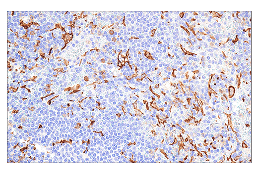
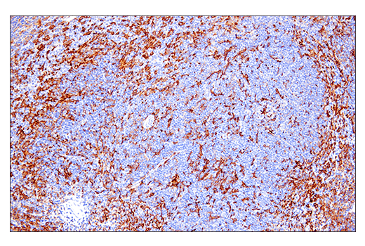
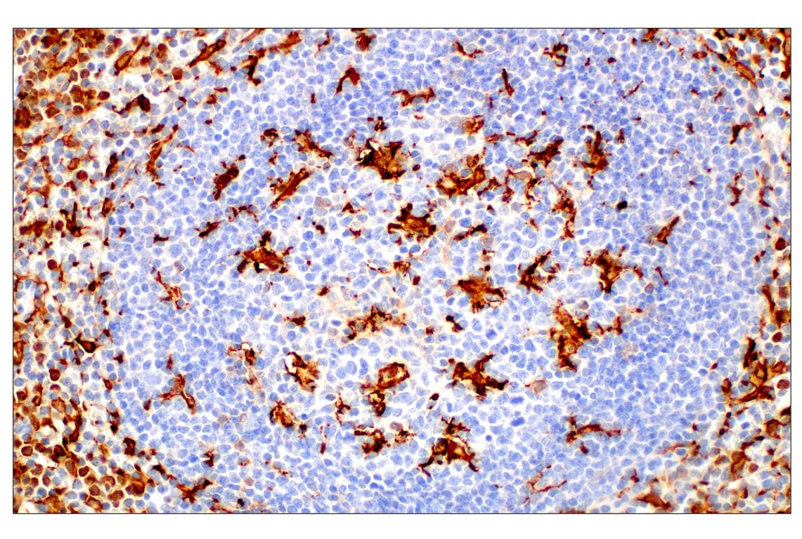
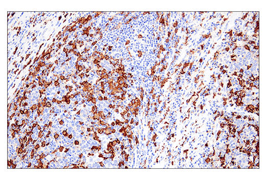
Immunohistochemical analysis of paraffin-embedded human non-Hodgkin's lymphoma using Iba1/AIF-1 (E4O4W) XP® Rabbit mAb performed on the Leica® BOND™ Rx.
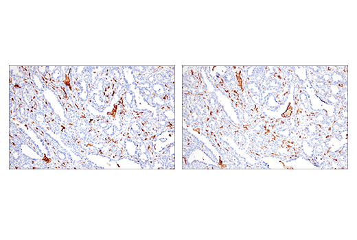
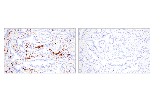
Immunohistochemical analysis of paraffin-embedded human ovarian clear cell carcinoma using Iba1/AIF-1 (E4O4W) XP® Rabbit mAb (left) or Iba1/AIF-1 (E5N4J) Mouse mAb (IHC Formulated) #58970 (right) performed on the Leica® BOND™ Rx. These two antibodies detect independent, unique epitopes on human Iba1/AIF-1 protein. The similar staining patterns obtained with both antibodies help to confirm the specificity of the staining.
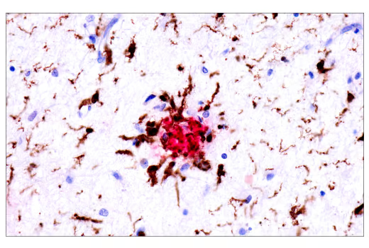
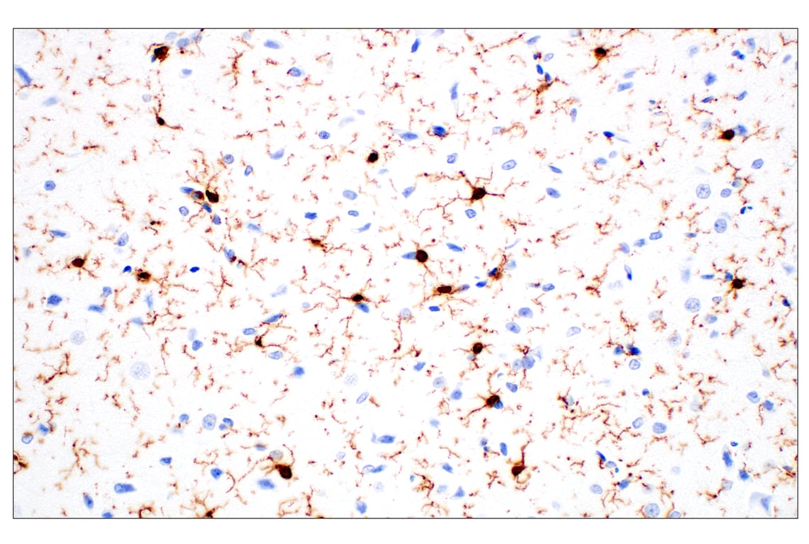
Dual immunohistochemical analysis of paraffin-embedded human Alzheimer's brain using Iba1/AIF-1 (E4O4W) XP® Rabbit mAb (brown) and APP/β-Amyloid (NAB228) Mouse mAb #2450 (red).
Immunohistochemical analysis of paraffin-embedded mouse brain using Iba1/AIF-1 (E4O4W) XP® Rabbit mAb.
Immunohistochemical analysis of paraffin-embedded mouse spleen using Iba1/AIF-1 (E4O4W) XP® Rabbit mAb.
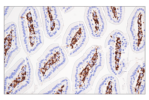
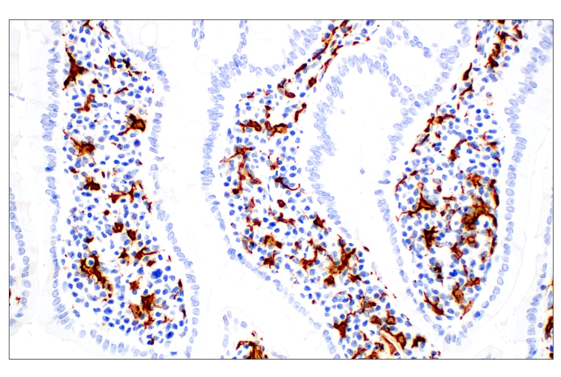
Immunohistochemical analysis of paraffin-embedded mouse small intestine using Iba1/AIF-1 (E4O4W) XP® Rabbit mAb.
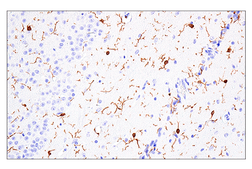
Immunohistochemical analysis of paraffin-embedded normal rat brain using Iba1/AIF-1 (E4O4W) XP® Rabbit mAb.
Immunohistochemical analysis of paraffin-embedded normal rhesus monkey spleen using Iba1/AIF-1 (E4O4W) XP® Rabbit mAb.
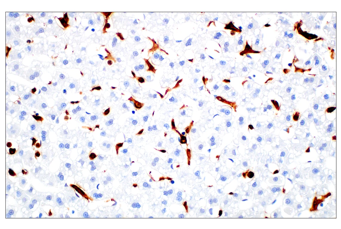
Immunohistochemical analysis of paraffin-embedded normal rhesus monkey liver using Iba1/AIF-1 (E4O4W) XP® Rabbit
Immunohistochemical analysis of paraffin-embedded normal Syrian hamster small intestine using Iba1/AIF-1 (E4O4W) XP® Rabbit mAb.
Immunohistochemical analysis of paraffin-embedded human ovarian serous carcinoma using Iba1/AIF-1 (E4O4W) XP® Rabbit mAb.
Immunohistochemical analysis of paraffin-embedded human colon carcinoma using Iba1/AIF-1 (E4O4W) XP® Rabbit mAb.
Immunohistochemical analysis of paraffin-embedded human ductal breast carcinoma using Iba1/AIF-1 (E4O4W) XP® Rabbit mAb (left) compared to concentration-matched Rabbit (DA1E) mAb IgG XP® Isotype Control #3900 (right).
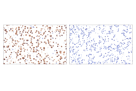
Immunohistochemical analysis of paraffin-embedded THP-1 cell pellet (left, positive) or SH-SY5Y cell pellet (right, negative) using Iba1/AIF-1 (E4O4W) XP® Rabbit mAb.
Confocal immunofluorescent analysis of human cortex (left) and mouse CA1 hippocampus (right) using Iba1/AIF-1 (E4O4W) XP® Rabbit mAb (green). In mouse tissue sections, cell nuclei were labeled with DAPI (blue). Images kindly provided by Dr. Simone Brioschi and Dr. Marco Colonna (Washington University) and used with permission.
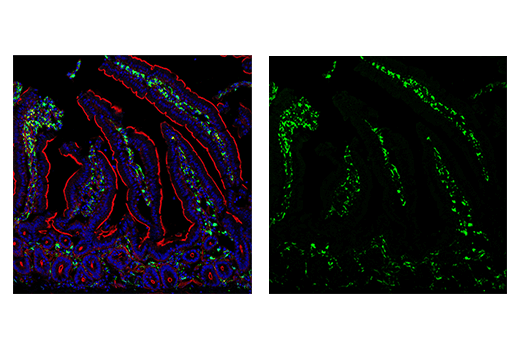
Confocal immunofluorescent analysis of mouse small intestine using Iba1/AIF-1 (E4O4W) XP® Rabbit mAb (green). Actin filaments were labeled with DyLight™ 554 Phalloidin #13054 (red). Sections were mounted in ProLong® Gold Antifade Reagent with DAPI #8961 (blue).
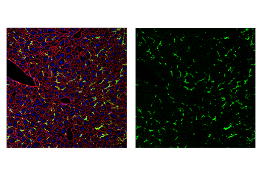
Confocal immunofluorescent analysis of mouse liver using Iba1/AIF-1 (E4O4W) XP® Rabbit mAb (green). Actin filaments were labeled with DyLight™ 554 Phalloidin #13054 (red). Sections were mounted in ProLong® Gold Antifade Reagent with DAPI #8961 (blue).
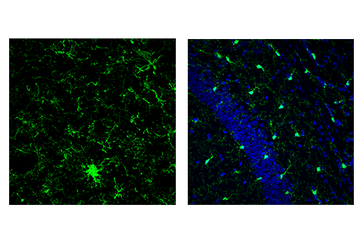
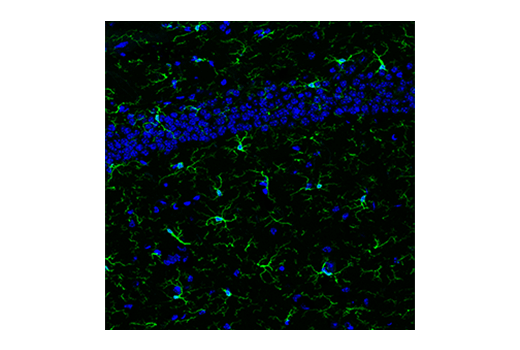
Confocal immunofluorescent analysis of microglia in mouse hippocampus using Iba1/AIF-1 (E4O4W) XP® Rabbit mAb (green). Sections were mounted in ProLong® Gold Antifade Reagent with DAPI #8961 (blue).
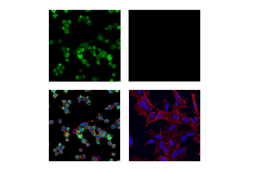
Confocal immunofluorescent analysis of THP-1 cells differentiated with TPA (12-O-Tetradecanoylphorbol-13-Acetate) #4174 (80 nM, 24 hr; left, positive) and SH-SY5Y cells (right, negative), using Iba1/AIF-1 (E4O4W) XP® Rabbit mAb (green). Actin filaments were labeled with DyLight™ 554 Phalloidin #13054 (red). Samples were mounted in ProLong® Gold Antifade Reagent with DAPI #8961 (blue).