


 全部商品分类


 用小程序,查商品更便捷


WB
1:5000-1:50000IP
1:50IHC-P
1:2000IF
1:500
Lamin-B1 is a protein that in humans is encoded by the LMNB1 gene. It is one of the essential members of the nuclear lamina protein family. Its main function is to maintain the integrity of nuclear skeleton, as well as to participate in the cell proliferation and aging by affecting the chromosome distribution. gene expression, and DNA damage repair. The abnormal expression of lamin B1 is related to certain diseases, including neurological diseases [e.g. neural tube defects (NDTs), adult-onset autosomal dominant leukodystrophy (ADLD)] and tumors (e.g. pancreatic cancer). It is also a potential tumor marker as well as drug target [PMID: 30499270].
12 months from date of receipt / reconstitution, -20 °C as supplied
参考图片
WB result of Lamin B1 Rabbit mAb Primary antibody: Lamin B1 Rabbit mAb at 1/1000 dilution Lane 1: HeLa whole cell lysate 20 µg Lane 2: Ramos whole cell lysate 20 µg Lane 3: MCF7 whole cell lysate 20 µg Lane 4: Jurkat whole cell lysate 20 µg Lane 5: MOLT-4 whole cell lysate 20 µg Lane 6: Caco-2 whole cell lysate 20 µg Secondary antibody: Goat Anti-Rabbit IgG, (H+L), HRP conjugated at 1/10000 dilution Predicted MW: 68 kDa Observed MW: 68 kDa
WB result of Lamin B1 Rabbit mAb Primary antibody: Lamin B1 Rabbit mAb at 1/1000 dilution Lane 1: NIH/3T3 whole cell lysate 20 µg Lane 2: RAW 264.7 whole cell lysate 20 µg Lane 3: mouse brain lysate 20 µg Lane 4: mouse heart lysate 20 µg Lane 5: mouse kidney lysate 20 µg Lane 6: mouse spleen lysate 20 µg Secondary antibody: Goat Anti-Rabbit IgG, (H+L), HRP conjugated at 1/10000 dilution Predicted MW: 68 kDa Observed MW: 68 kDa
WB result of Lamin B1 Rabbit mAb Primary antibody: Lamin B1 Rabbit mAb at 1/1000 dilution Lane 1: PC-12 whole cell lysate 20 µg Lane 2: rat brain lysate 20 µg Lane 3: rat heart lysate 20 µg Lane 4: rat kidney lysate 20 µg Secondary antibody: Goat Anti-Rabbit IgG, (H+L), HRP conjugated at 1/10000 dilution Predicted MW: 68 kDa Observed MW: 68 kDa
IHC shows positive staining in paraffin-embedded human spleen. Anti-Lamin B1 antibody was used at 1/2000 dilution, followed by a Goat Anti-Rabbit IgG H&L (HRP) ready to use. Counterstained with hematoxylin. Heat mediated antigen retrieval with Tris/EDTA buffer pH9.0 was performed before commencing with IHC staining protocol.
IHC shows positive staining in paraffin-embedded human stomach. Anti-Lamin B1 antibody was used at 1/2000 dilution, followed by a Goat Anti-Rabbit IgG H&L (HRP) ready to use. Counterstained with hematoxylin. Heat mediated antigen retrieval with Tris/EDTA buffer pH9.0 was performed before commencing with IHC staining protocol.
IHC shows positive staining in paraffin-embedded human endometrial cancer. Anti-Lamin B1 antibody was used at 1/2000 dilution, followed by a Goat Anti-Rabbit IgG H&L (HRP) ready to use. Counterstained with hematoxylin. Heat mediated antigen retrieval with Tris/EDTA buffer pH9.0 was performed before commencing with IHC staining protocol.
IHC shows positive staining in paraffin-embedded human lung cancer. Anti-Lamin B1 antibody was used at 1/2000 dilution, followed by a Goat Anti-Rabbit IgG H&L (HRP) ready to use. Counterstained with hematoxylin. Heat mediated antigen retrieval with Tris/EDTA buffer pH9.0 was performed before commencing with IHC staining protocol.
IHC shows positive staining in paraffin-embedded human thyroid cancer. Anti-Lamin B1 antibody was used at 1/2000 dilution, followed by a Goat Anti-Rabbit IgG H&L (HRP) ready to use. Counterstained with hematoxylin. Heat mediated antigen retrieval with Tris/EDTA buffer pH9.0 was performed before commencing with IHC staining protocol.
IHC shows positive staining in paraffin-embedded mouse spleen. Anti-Lamin B1 antibody was used at 1/2000 dilution, followed by a Goat Anti-Rabbit IgG H&L (HRP) ready to use. Counterstained with hematoxylin. Heat mediated antigen retrieval with Tris/EDTA buffer pH9.0 was performed before commencing with IHC staining protocol.
IHC shows positive staining in paraffin-embedded mouse skeletal muscle. Anti-Lamin B1 antibody was used at 1/2000 dilution, followed by a Goat Anti-Rabbit IgG H&L (HRP) ready to use. Counterstained with hematoxylin. Heat mediated antigen retrieval with Tris/EDTA buffer pH9.0 was performed before commencing with IHC staining protocol.
IHC shows positive staining in paraffin-embedded rat cerebral cortex. Anti-Lamin B1 antibody was used at 1/2000 dilution, followed by a Goat Anti-Rabbit IgG H&L (HRP) ready to use. Counterstained with hematoxylin. Heat mediated antigen retrieval with Tris/EDTA buffer pH9.0 was performed before commencing with IHC staining protocol.
IHC shows positive staining in paraffin-embedded rat liver. Anti-Lamin B1 antibody was used at 1/2000 dilution, followed by a Goat Anti-Rabbit IgG H&L (HRP) ready to use. Counterstained with hematoxylin. Heat mediated antigen retrieval with Tris/EDTA buffer pH9.0 was performed before commencing with IHC staining protocol.
Lamin B1 Rabbit mAb at 1/50 dilution (1 µg) immunoprecipitating Lamin B1 in 0.4 mg HeLa whole cell lysate.
Western blot was performed on the immunoprecipitate using Lamin B1 Rabbit mAb at 1/1000 dilution.
Secondary antibody (HRP) for IP was used at 1/400 dilution.
Lane 1: HeLa whole cell lysate 20 µg (Input)
Lane 2: Lamin B1 Rabbit mAb IP in HeLa whole cell lysate
Lane 3: Rabbit monoclonal IgG IP in HeLa whole cell lysate
Predicted MW: 68 kDa
Observed MW: 68 kDa
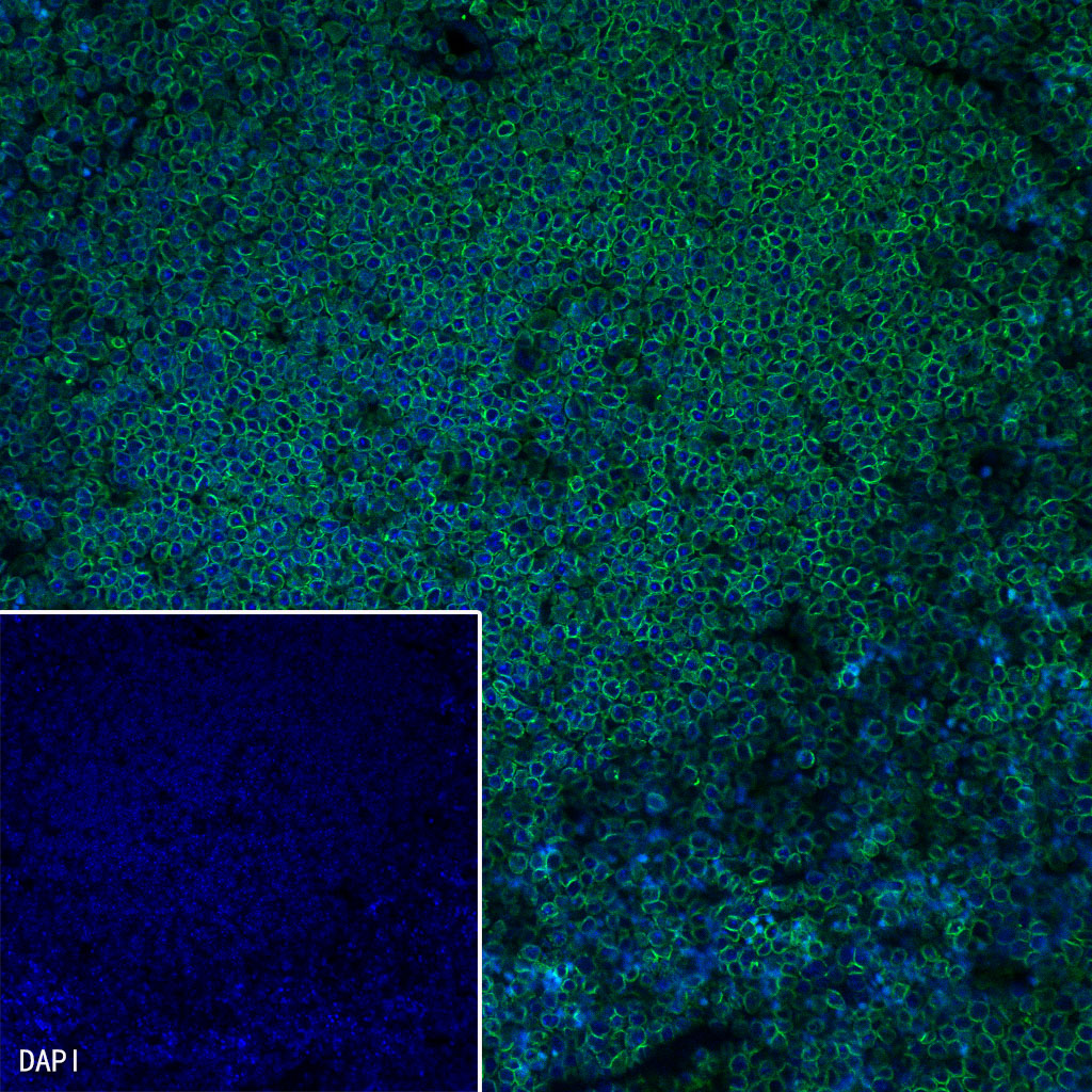
IF shows positive staining in paraffin-embedded mouse spleen. Anti-Lamin B1 antibody was used at 1/500 dilution (Green) and incubated overnight at 4°C. Goat polyclonal Antibody to Rabbit IgG - H&L (Alexa Fluor® 488) was used as secondary antibody at 1/1000 dilution. Counterstained with DAPI (Blue). Heat mediated antigen retrieval with EDTA buffer pH9.0 was performed before commencing with IF staining protocol.
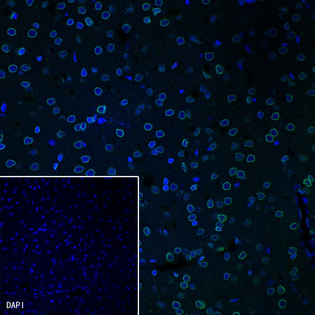
IF shows positive staining in paraffin-embedded rat brain. Anti-Lamin B1 antibody was used at 1/500 dilution (Green) and incubated overnight at 4°C. Goat polyclonal Antibody to Rabbit IgG - H&L (Alexa Fluor® 488) was used as secondary antibody at 1/1000 dilution. Counterstained with DAPI (Blue). Heat mediated antigen retrieval with EDTA buffer pH9.0 was performed before commencing with IF staining protocol.