Human (QC Testing), Rhesus, Cynomolgus, Baboon (Tested in Development)
来源宿主:
Mouse BALB/c IgG1, κ
展开
产品介绍
产品介绍
产品信息
抗原名称
IFN-γ
宿主
Mouse BALB/c IgG1, κ
免疫原
Human IFN-γ from supernatants of S. aureus-stimulated PBMC
简单描述
The 4S.B3 monoclonal antibody specifically binds to interferon-γ (IFN-γ). The immunogen used to generate this hybridoma was partially purified human IFN-γ obtained from supernatants of human PBMC stimulated with
Staphylococcus aureus
. Interferon-γ (IFN-γ) is a potent multifunctional cytokine that is produced by several activated cell types including NK, NKT, CD4+TCRαβ+, CD8+TCRαβ+, and TCRγδ+ T cells. IFN-γ exerts its biological effects through specific binding to the high-affinity IFN-γ Receptor Complex comprised of IFN-γRα (CD119) and IFN-γRβ subunits. In addition to its antiviral effects, IFN-γ upregulates a number of lymphoid cell functions including the antimicrobial and antitumor responses of macrophages, NK cells, and neutrophils. In addition, IFN-γ can exert strong regulatory influences on the proliferation, differentiation, and effector responses of B cell and T cell subsets. These influences can involve IFN-γ's capacity to boost MHC class I and II expression by antigen-presenting cells as well as to direct effects on B cells and T cells themselves. Human IFN-γ is a 14-18 kDa glycoprotein containing 143 amino acid residues.
Clone 4S.B3 also cross-reacts with a cytoplasmic component of peripheral blood CD3+ lymphocytes of baboon, and both rhesus and cynomolgus macaque monkeys following five-hour treatment with phorbol myristic acetate (PMA) and Ca++ ionophore (A23187) in the presence of monensin. The staining pattern of 4S.B3 in CD3+ cells is similar to that observed with peripheral blood T lymphocytes from normal human donors. This reagent is useful for intracellular immunofluorescent staining for flow cytometric analysis to identify and enumerate IFN-γ + cells within a mixed cell population.
商品描述
4S.B3
The 4S.B3 monoclonal antibody specifically binds to interferon-γ (IFN-γ). The immunogen used to generate this hybridoma was partially purified human IFN-γ obtained from supernatants of human PBMC stimulated with
Staphylococcus aureus
. Interferon-γ (IFN-γ) is a potent multifunctional cytokine that is produced by several activated cell types including NK, NKT, CD4+TCRαβ+, CD8+TCRαβ+, and TCRγδ+ T cells. IFN-γ exerts its biological effects through specific binding to the high-affinity IFN-γ Receptor Complex comprised of IFN-γRα (CD119) and IFN-γRβ subunits. In addition to its antiviral effects, IFN-γ upregulates a number of lymphoid cell functions including the antimicrobial and antitumor responses of macrophages, NK cells, and neutrophils. In addition, IFN-γ can exert strong regulatory influences on the proliferation, differentiation, and effector responses of B cell and T cell subsets. These influences can involve IFN-γ's capacity to boost MHC class I and II expression by antigen-presenting cells as well as to direct effects on B cells and T cells themselves. Human IFN-γ is a 14-18 kDa glycoprotein containing 143 amino acid residues.
Clone 4S.B3 also cross-reacts with a cytoplasmic component of peripheral blood CD3+ lymphocytes of baboon, and both rhesus and cynomolgus macaque monkeys following five-hour treatment with phorbol myristic acetate (PMA) and Ca++ ionophore (A23187) in the presence of monensin. The staining pattern of 4S.B3 in CD3+ cells is similar to that observed with peripheral blood T lymphocytes from normal human donors. This reagent is useful for intracellular immunofluorescent staining for flow cytometric analysis to identify and enumerate IFN-γ + cells within a mixed cell population.
同种型
Mouse BALB/c IgG1, κ
克隆号
克隆 4S.B3 (RUO)
浓度
0.5 mg/ml
产品详情
Purified
Tissue culture supernatant is purified by either protein A/G or affinity purification methods. Both methods yield antibody in solution that is free of most other soluble proteins, lipids, etc. This format provides pure antibody that is suitable for a number of downstream applications including: secondary labeling for flow cytometry or microscopy, ELISA, Western blot, etc.
应用
实验应用
Intracellular block/flow cytometry (Routinely Tested), ELISA Detection, Western blot (Reported)
反应种属
Human (QC Testing), Rhesus, Cynomolgus, Baboon (Tested in Development)
目标/特异性
IFN-γ
背景
别名
IFNG; Interferon-gamma; IFG; IFI; Type II interferon
研发参考(3)
1. Meager A, Parti S, Barwick S, Spragg J, O'Hagan K. Detection of hybridomas secreting monoclonal antibodies to human gamma interferon using a rapid screening technique and specificity of certain monoclonal antibodies to gamma interferon. J Interferon Res. 1984; 4(4):619-625. (Biology).
2. Meager A. Characterization of interferons and immunoassays. In: Clemens MJ, Morris AG, Gearing AJH, ed. Lymphokines and Interferons. A Practical Approach. Oxford: IRL Press Ltd; 1987:105-127.
3. Prussin C, Metcalfe DD. Detection of intracytoplasmic cytokine using flow cytometry and directly conjugated anti-cytokine antibodies. J Immunol Methods. 1995; 188(1):117-128. (Methodology).
数据库链接
Entrez-Gene ID
3458
参考图片
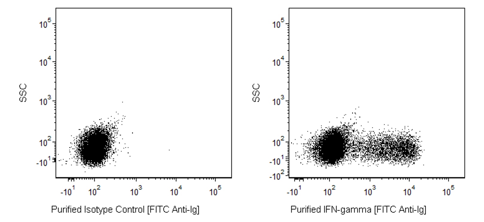
Flow cytometric analysis of IFN-γ expression in stimulated human peripheral blood mononuclear cells. HiCK-1 Human Cytokine Positive Control Cells (Cat. No. 555061) were permeabilized with BD Perm/Wash™ Buffer (Cat. No. 554723). The cells were then stained with either Purified Mouse IgG1, κ Isotype Control (Cat No. 556648, Left Panel) or with the Purified Mouse Anti-Human IFN-γ antibody (Cat No. 554549, Right Panel), followed by FITC Goat Anti-Mouse Ig (Cat. No. 554001). Flow cytometric dot plots showing the expression of IFN-γ (or Ig Isotype Control staining) versus side scatter were derived from gated events with the forward and side light-scatter characteristics of intact lymphocytes. Flow cytometric analysis was performed using a BD FACSCanto™ System.
Flow cytometric analysis of IFN-γ expression in stimulated human peripheral blood mononuclear cells. HiCK-1 Human Cytokine Positive Control Cells (Cat. No. 555061) were permeabilized with BD Perm/Wash™ Buffer (Cat. No. 554723). The cells were then stained with either Purified Mouse IgG1, κ Isotype Control (Cat No. 556648, Left Panel) or with the Purified Mouse Anti-Human IFN-γ antibody (Cat No. 554549, Right Panel), followed by FITC Goat Anti-Mouse Ig (Cat. No. 554001). Flow cytometric dot plots showing the expression of IFN-γ (or Ig Isotype Control staining) versus side scatter were derived from gated events with the forward and side light-scatter characteristics of intact lymphocytes. Flow cytometric analysis was performed using a BD FACSCanto™ System.


 全部商品分类


 下载产品说明书
 用小程序,查商品更便捷


 收藏
收藏
 对比
对比 咨询
咨询