The XMG1.2 monoclonal antibody specifically binds to mouse interferon-γ (IFN-γ) protein. IFN-γ is a pleiotropic cytokine, of approximately 15-17 kDa, involved in the regulation of inflammatory and immune responses. It plays an important role in activation, growth, and differentiation of T and B lymphocytes, macrophages, NK cells and other non-hematopoietic cell types. IFN-γ production is associated with the Th1 cell differentiation. The purified form of this antibody has been reported to be a neutralizing antibody.
商品描述
XMG1.2
The XMG1.2 monoclonal antibody specifically binds to mouse interferon-γ (IFN-γ) protein. IFN-γ is a pleiotropic cytokine, of approximately 15-17 kDa, involved in the regulation of inflammatory and immune responses. It plays an important role in activation, growth, and differentiation of T and B lymphocytes, macrophages, NK cells and other non-hematopoietic cell types. IFN-γ production is associated with the Th1 cell differentiation. The purified form of this antibody has been reported to be a neutralizing antibody.
同种型
Rat IgG1, κ
克隆号
克隆 XMG1.2 (RUO)
浓度
0.2 mg/ml
产品详情
PE-Cy7
PE-Cy7 dye is a part of the BD PE family of dyes. This tandem fluorochrome is comprised of a R-Phycoerythrin (PE) donor that has excitation maxima (Ex Max) of 496-nm and 566-nm and an acceptor dye, Cy™7, with an emission maximum (Em Max) at 781-nm. PE can be excited by the Blue (488-nm), Green (532-nm) and yellow-green (561-nm) lasers and detected using an optical filter centered near 781 nm (e.g., a 760/60-nm bandpass filter). The donor dye can be excited by the Blue (488-nm), Green (532-nm) and yellow-green (561-nm) lasers and the acceptor dye can be excited by the Red (627–640-nm) laser resulting in cross-laser excitation and fluorescence spillover. Please ensure that your instrument’s configurations (lasers and optical filters) are appropriate for this dye.
研发参考(4)
1. Abrams JS, Roncarolo MG, Yssel H, Andersson U, Gleich GJ, Silver JE. Strategies of anti-cytokine monoclonal antibody development: immunoassay of IL-10 and IL-5 in clinical samples. Immunol Rev. 1992; 127:5-24. (Clone-specific).
2. Cherwinski HM, Schumacher JH, Brown KD, Mosmann TR. Two types of mouse helper T cell clone. III. Further differences in lymphokine synthesis between Th1 and Th2 clones revealed by RNA hybridization, functionally monospecific bioassays, and monoclonal antibodies. J Exp Med. 1987; 166(5):1229-1244. (Clone-specific).
3. Prussin C, Metcalfe DD. Detection of intracytoplasmic cytokine using flow cytometry and directly conjugated anti-cytokine antibodies. J Immunol Methods. 1995; 188(1):117-128. (Methodology: Flow cytometry).
4. Sander B, Hoiden I, Andersson U, Moller E, Abrams JS. Similar frequencies and kinetics of cytokine producing cells in murine peripheral blood and spleen. Cytokine detection by immunoassay and intracellular immunostaining. J Immunol Methods. 1993; 166(2):201-214. (Clone-specific).
数据库链接
Entrez-Gene ID
15978
参考图片
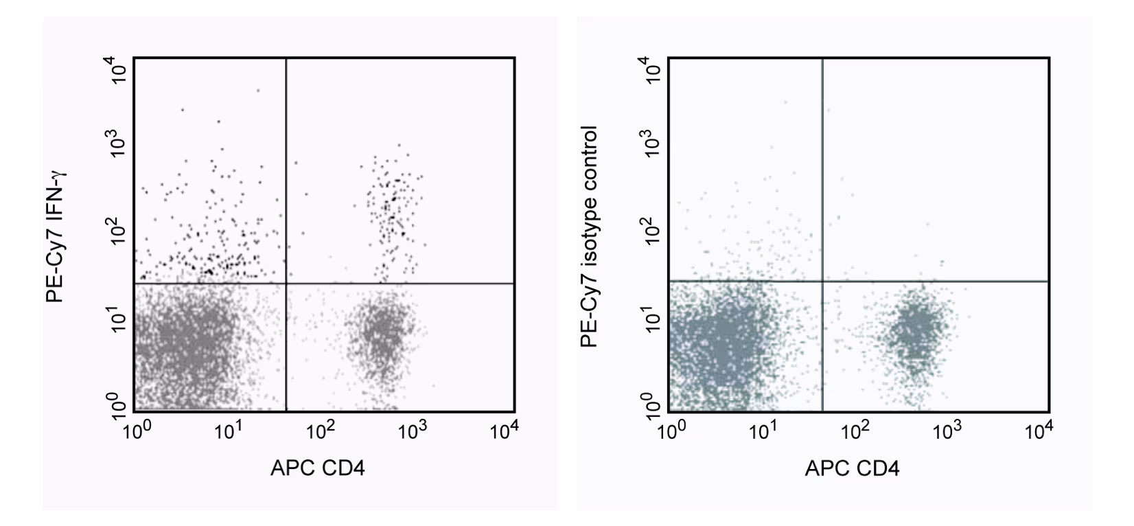
Expression of IFN- by stimulated CD4+ and CD4-BALB/c spleen cells. Splenocytes from BALB/C mice were stimulated for 4 hrs with PMA (5 ng/ml, Sigma, Cat. No. P-8139) and Ionomycin (500 µg/ml, Sigma Cat. No. I-0634) in the presence of Brefeldin A (GolgiPlug, Cat. No. 555029). Cells were harvested, fixed, permeabilized and stained with APC rat anti-mouse CD4 (APC-RM4-5, Cat. No. 553051) and either rat anti-mouse IFN-γ antibody (PE-Cy7-XMG1.2, Cat. No. 557649), (left panel) or immunoglobulin isotype control (PE-Cy7-R3-34, Cat. No. 557645), (right panel) by using the BD Pharmingen staining protocol. To demonstrate specificity of staining the binding of PE-Cy7-XMG1.2 was blocked by preincubation of the fixed/permeabilized cells with an excess of unlabelled XMG1.2 antibody (5 µg, Cat. No. 554409, data not shown) prior to stainining. The quadarant markers for the bivariate dot plots were set based on the autofluorescence and isotype controls.
Expression of IFN- by stimulated CD4+ and CD4-BALB/c spleen cells. Splenocytes from BALB/C mice were stimulated for 4 hrs with PMA (5 ng/ml, Sigma, Cat. No. P-8139) and Ionomycin (500 µg/ml, Sigma Cat. No. I-0634) in the presence of Brefeldin A (GolgiPlug, Cat. No. 555029). Cells were harvested, fixed, permeabilized and stained with APC rat anti-mouse CD4 (APC-RM4-5, Cat. No. 553051) and either rat anti-mouse IFN-γ antibody (PE-Cy7-XMG1.2, Cat. No. 557649), (left panel) or immunoglobulin isotype control (PE-Cy7-R3-34, Cat. No. 557645), (right panel) by using the BD Pharmingen staining protocol. To demonstrate specificity of staining the binding of PE-Cy7-XMG1.2 was blocked by preincubation of the fixed/permeabilized cells with an excess of unlabelled XMG1.2 antibody (5 µg, Cat. No. 554409, data not shown) prior to stainining. The quadarant markers for the bivariate dot plots were set based on the autofluorescence and isotype controls.


 全部商品分类


 下载产品说明书
 用小程序,查商品更便捷


 收藏
收藏
 对比
对比 咨询
咨询
