The B27 monoclonal antibody specifically binds to human interferon-γ (IFN-γ), a 14-18 kDa glycoprotein containing 143 amino acid residues. IFN-γ is a potent multifunctional cytokine produced by several activated cell types including NK, NKT, CD4+TCRαβ+, CD8+TCRαβ+, and TCRγδ+ T cells. IFN-γ exerts its biological effects through specific binding to the high-affinity IFN-γ receptor complex comprised of IFN-γRα (CD119) and IFN-γRβ subunits. In addition to its antiviral effects, IFN-γ upregulates a number of lymphoid cell functions including the antimicrobial and anti-tumor responses of macrophages, NK cells, and neutrophils. In addition, IFN-γ influences the regulation of proliferation, differentiation, and effector responses of B cell and T cell subsets. These influences can involve IFN-γ's capacity to boost MHC class I and II expression by antigen-presenting cells as well as direct effects on B cells and T cells themselves. B27 is a neutralizing antibody. The use of B27 antibody for epitope mapping of human IFN-γ has been described. The B27 antibody has been reported not to bind to denatured IFN-γ.
商品描述
B27
The B27 monoclonal antibody specifically binds to human interferon-γ (IFN-γ), a 14-18 kDa glycoprotein containing 143 amino acid residues. IFN-γ is a potent multifunctional cytokine produced by several activated cell types including NK, NKT, CD4+TCRαβ+, CD8+TCRαβ+, and TCRγδ+ T cells. IFN-γ exerts its biological effects through specific binding to the high-affinity IFN-γ receptor complex comprised of IFN-γRα (CD119) and IFN-γRβ subunits. In addition to its antiviral effects, IFN-γ upregulates a number of lymphoid cell functions including the antimicrobial and anti-tumor responses of macrophages, NK cells, and neutrophils. In addition, IFN-γ influences the regulation of proliferation, differentiation, and effector responses of B cell and T cell subsets. These influences can involve IFN-γ's capacity to boost MHC class I and II expression by antigen-presenting cells as well as direct effects on B cells and T cells themselves. B27 is a neutralizing antibody. The use of B27 antibody for epitope mapping of human IFN-γ has been described. The B27 antibody has been reported not to bind to denatured IFN-γ.
同种型
Mouse IgG1, κ
克隆号
克隆 B27 (RUO)
产品详情
PE
R-Phycoerythrin (PE), is part of the BD family of Phycobiliprotein dyes. This fluorochrome is a multimeric fluorescent phycobiliprotein with excitation maximum (Ex Max) of 496 nm and 566 nm and an emission maximum (Em Max) at 576 nm. PE is designed to be excited by the Blue (488 nm), Green (532 nm) and Yellow-Green (561 nm) lasers and detected using an optical filter centered near 575 nm (e.g., a 575/26-nm bandpass filter). As PE is excited by multiple lasers, this can result in cross-laser excitation and fluorescence spillover on instruments with various combinations of Blue, Green, and Yellow-Green lasers. Please ensure that your instrument’s configurations (lasers and optical filters) are appropriate for this dye.
Human (QC Testing), Rhesus, Cynomolgus, Baboon (Tested in Development)
目标/特异性
IFN-γ
背景
别名
IFNG; Interferon-gamma; Interferon-γ; Type II interferon; MAF
制备和贮存
存储溶液
Aqueous buffered solution containing BSA and ≤0.09% sodium azide.
保存方式
Aqueous buffered solution containing BSA and ≤0.09% sodium azide.
文献
文献
研发参考(5)
1. Abrams JS, Roncarolo MG, Yssel H, Andersson U, Gleich GJ, Silver JE. Strategies of anti-cytokine monoclonal antibody development: immunoassay of IL-10 and IL-5 in clinical samples. Immunol Rev. 1992; 127:5-24. (Clone-specific).
2. Favre C, Wijdenes J, Cabrillat H, Djossou O, Banchereau J, de Vries JE. Epitope mapping of recombinant human gamma interferon using monoclonal antibodies. Mol Immunol. 1989; 26(1):17-25. (Clone-specific: Immunoprecipitation, Neutralization).
3. Fonteneau JF, Le Drean E, Le Guiner S, Gervois N, Diez E, Jotereau F. Heterogeneity of biologic responses of melanoma-specific CTL. J Immunol. 1997; 159(6):2831-2839. (Biology).
4. Prussin C, Metcalfe DD. Detection of intracytoplasmic cytokine using flow cytometry and directly conjugated anti-cytokine antibodies. J Immunol Methods. 1995; 188(1):117-128. (Methodology: Flow cytometry).
5. Rotteveel FT, Kokkelink I, van Lier RA, et al. Clonal analysis of functionally distinct human CD4+ T cell subsets. J Exp Med. 1988; 168(5):1659-1673. (Biology).
数据库链接
Entrez-Gene ID
15978,16176,25712,3458,380793,4049
参考图片
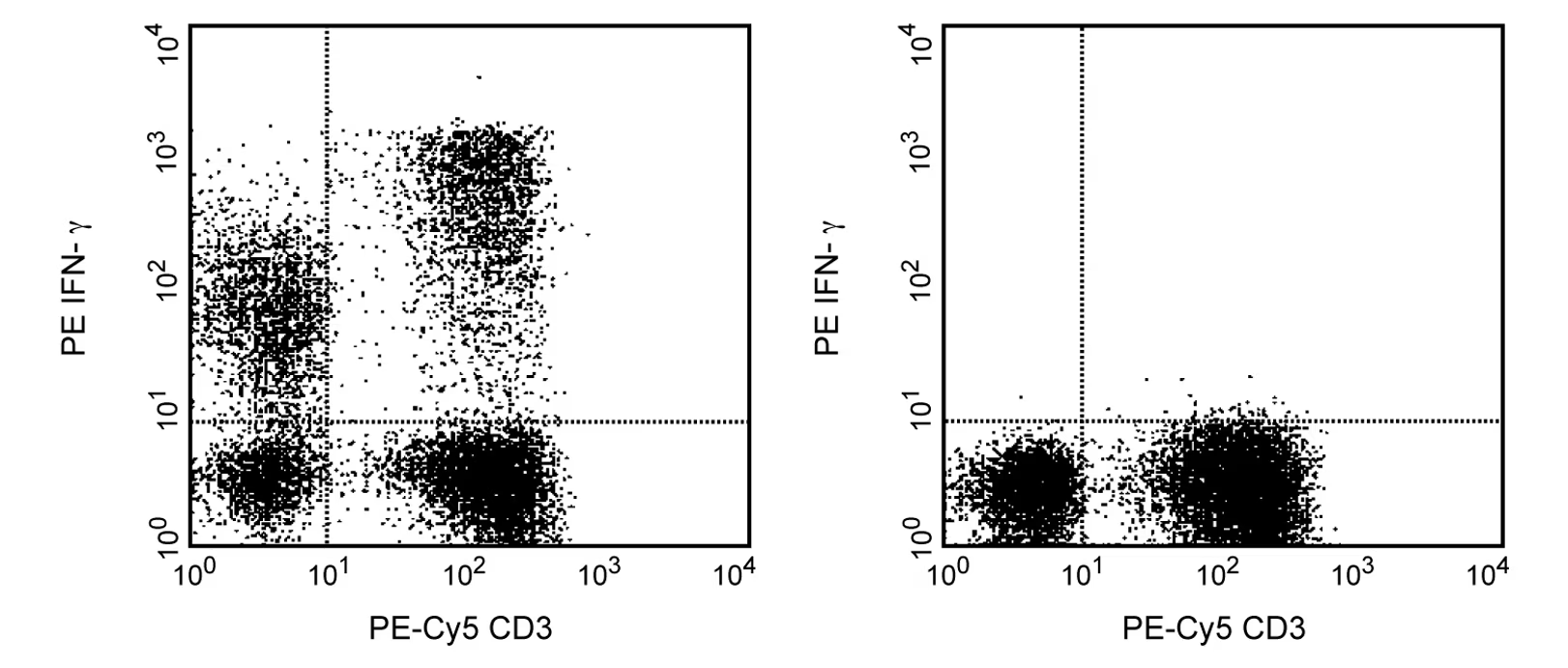
Expression of IFN-γ by stimulated human peripheral blood mononuclear cells (PBMC). Human PBMC were stimulated for 6 h with PMA (50 ng/ml; Sigma, Cat. #P-8139) and calcium ionophore A23187 (250 ng/ml; Sigma, Cat. #C-9275) in the presence of GolgiStop™ (2 µM final concentration; Cat. No. 554715). The PBMC were stained with PE-Cy™5 Mouse Anti-Human CD3 (Cat. 555334), fixed, permeabilized, and subsequently stained with 20 µl of PE Mouse Anti-Human IFN-γ (Cat. No. 559327/554701; left panel). To demonstrate specificity of staining, binding by the PE-B27 antibody was blocked by preincubation of fixed/permeabilized cells with Purified Mouse Anti-Human IFN-γ (5 µg; Cat. No. 554699/550011; right panel) prior to staining. The quadrant markers for the bivariate dot plot were set based on autofluorescence controls and verified using the unlabeled antibody blocking control.
Expression of IFN-γ by stimulated human peripheral blood mononuclear cells (PBMC). Human PBMC were stimulated for 6 h with PMA (50 ng/ml; Sigma, Cat. #P-8139) and calcium ionophore A23187 (250 ng/ml; Sigma, Cat. #C-9275) in the presence of GolgiStop™ (2 µM final concentration; Cat. No. 554715). The PBMC were stained with PE-Cy™5 Mouse Anti-Human CD3 (Cat. 555334), fixed, permeabilized, and subsequently stained with 20 µl of PE Mouse Anti-Human IFN-γ (Cat. No. 559327/554701; left panel). To demonstrate specificity of staining, binding by the PE-B27 antibody was blocked by preincubation of fixed/permeabilized cells with Purified Mouse Anti-Human IFN-γ (5 µg; Cat. No. 554699/550011; right panel) prior to staining. The quadrant markers for the bivariate dot plot were set based on autofluorescence controls and verified using the unlabeled antibody blocking control.


 全部商品分类


 下载产品说明书
 用小程序,查商品更便捷


 收藏
收藏
 对比
对比 咨询
咨询
