


 全部商品分类


BD Horizon™ BV650 Rat Anti-Mouse IFN-γ


 下载产品说明书 下载SDS
 用小程序,查商品更便捷


 收藏
收藏
 对比
对比 咨询
咨询

参考图片
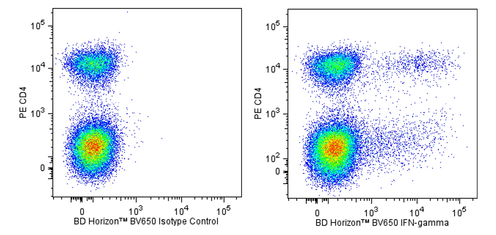
Two-color flow cytometric analysis of IFN-γ expression by stimulated mouse splenocytes. Mouse splenic leucocytes were stimulated for 5 hours with Phorbol 12-Myristate 13-Acetate (PMA; Sigma P-8139; 50 ng/ml) and Ionomycin (Sigma I-0634; 1 μg/ml) in the presence of BD GolgiStop™ Protein Transport Inhibitor (containing Monensin) (Cat. No. 554724). The cells were harvested, washed with BD Pharmingen™ Stain Buffer (FBS) (Cat. No. 554656), and fixed with BD Cytofix™ Fixation Buffer (Cat. No. 554655). The cells were then washed and stained in BD Perm/Wash™ Buffer (Cat. No. 554723) with PE Rat Anti-Mouse CD4 antibody (Cat. No. 553048/553049/561837) and either BD Horizon™ BV650 Rat IgG1, κ Isotype Control (Cat. No. 563848; Left Panel) or BD Horizon™ BV650 Rat Anti-Mouse IFN-γ antibody (Cat. No. 563854; Right Panel) by using the BD Biosciences Intracellular Cytokine Staining protocol. Two-color flow cytometric dot plots showing the correlated expression of IFN-γ (or Ig Isotype control staining) versus CD4 were derived from gated events with the forward and side light-scatter characteristics of intact stimulated leucocytes. Flow cytometric analysis was performed using a BD™ LSR II Flow Cytometer System.
Two-color flow cytometric analysis of IFN-γ expression by stimulated mouse splenocytes. Mouse splenic leucocytes were stimulated for 5 hours with Phorbol 12-Myristate 13-Acetate (PMA; Sigma P-8139; 50 ng/ml) and Ionomycin (Sigma I-0634; 1 μg/ml) in the presence of BD GolgiStop™ Protein Transport Inhibitor (containing Monensin) (Cat. No. 554724). The cells were harvested, washed with BD Pharmingen™ Stain Buffer (FBS) (Cat. No. 554656), and fixed with BD Cytofix™ Fixation Buffer (Cat. No. 554655). The cells were then washed and stained in BD Perm/Wash™ Buffer (Cat. No. 554723) with PE Rat Anti-Mouse CD4 antibody (Cat. No. 553048/553049/561837) and either BD Horizon™ BV650 Rat IgG1, κ Isotype Control (Cat. No. 563848; Left Panel) or BD Horizon™ BV650 Rat Anti-Mouse IFN-γ antibody (Cat. No. 563854; Right Panel) by using the BD Biosciences Intracellular Cytokine Staining protocol. Two-color flow cytometric dot plots showing the correlated expression of IFN-γ (or Ig Isotype control staining) versus CD4 were derived from gated events with the forward and side light-scatter characteristics of intact stimulated leucocytes. Flow cytometric analysis was performed using a BD™ LSR II Flow Cytometer System.