 全部商品分类

品牌: BD Pharmingen
 下载产品说明书
 下载SDS
下载SDS 用小程序,查商品更便捷


 收藏
收藏
 对比
对比 咨询
咨询反应种属:
Human (Tested in Development)
来源宿主:
Mouse BALB/c IgG1, λ
产品介绍
产品介绍
产品信息
荧光素标记
宿主
Mouse BALB/c IgG1, λ

免疫原
Human IgA1-λ myeloma Protein

简单描述
The 1-155-2 monoclonal antibody specifically recognizes human immunoglobulin lambda light chains (Igλ). Immunoglobulins are expressed on the surface of B lymphocytes and plasma cells, and they also circulate in the serum. The two light chains in an immunoglobulin are of the same type, either kappa (Igκ) or Igλ. Immunoglobulins bearing Igλ are expressed on a minority of normal B lymphocytes and on neoplastic cells of some leukemias, lymphomas, and plasmacytomas. In serum, the 1-155-2 antibody binds to immunoglobulins bearing Igλ as well as free Igλ. It does not bind to Igκ or immunoglobulin heavy chains.

商品描述
1-155-2
The 1-155-2 monoclonal antibody specifically recognizes human immunoglobulin lambda light chains (Igλ). Immunoglobulins are expressed on the surface of B lymphocytes and plasma cells, and they also circulate in the serum. The two light chains in an immunoglobulin are of the same type, either kappa (Igκ) or Igλ. Immunoglobulins bearing Igλ are expressed on a minority of normal B lymphocytes and on neoplastic cells of some leukemias, lymphomas, and plasmacytomas. In serum, the 1-155-2 antibody binds to immunoglobulins bearing Igλ as well as free Igλ. It does not bind to Igκ or immunoglobulin heavy chains.

同种型
Mouse BALB/c IgG1, λ

克隆号
克隆 1-155-2 (RUO)

浓度
0.2 mg/ml

产品详情
RY586
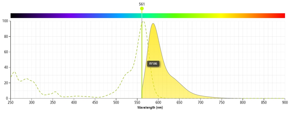
The BD Horizon RealYellow™ 586 (RY586) Dye is part of the BD family of yellow-green dyes. It is a small organic fluorochrome with an excitation maximum (Ex Max) at 565-nm and an emission maximum (Em Max) at 586-nm. Driven by BD innovation, RY586 can be used on both spectral and conventional cytometers and is designed to be excited by the Yellow-Green laser (561-nm) with minimal excitation by the 488-nm Blue laser. For conventional instruments equipped with a Yellow-Green laser (561-nm), RY586 can be used as an alternative to PE and we recommend using an optical filter centered near 586-nm (eg, a 586/15-nm bandpass filter). For spectral instruments equipped with a Yellow-Green laser (561-nm), it can be used in conjunction with PE. Compared to PE, RY586 is similar in brightness, minimal spillover into Blue detectors, and increased spillover into the 610/20-nm (PE-CF594) detector. Please ensure that your instrument configuration (lasers and optical filters) is appropriate for this dye.

RY586
Yellow-Green 561 nm
564 nm
586 nm
应用
实验应用
Flow cytometry (Qualified)

反应种属
Human (Tested in Development)

目标/特异性
Ig, λ Light Chain

背景
别名
IGL; IGL@; Ig λ; Immunoglobulin lambda

制备和贮存
存储溶液
Aqueous buffered solution containing ≤0.09% sodium azide.

保存方式
Aqueous buffered solution containing ≤0.09% sodium azide.
文献
文献
研发参考(13)
1. Ault KA. Flow cytometric evaluation of normal and neoplastic B cells. In: Rose NR, Friedman H, Fahey JL. Rose NR, Friedman H, Fahey JL, ed. Manual of Clinical Laboratory Immunology. 3rd ed.. Washington, DC: American Society for Microbiology; 1986:247-253.
2. Foon KA, Todd RF. Immunologic classification of leukemia and lymphoma.. Blood. 1986; 68(1):1-31. (Methodology).
3. Harris NL, Data RE. The distribution of neoplastic and normal B-lymphoid cells in nodular lymphomas: use of an immunoperoxidase technique on frozen sections.. Hum Pathol. 1982; 13(7):610-7. (Methodology: Immunohistochemistry).
4. Kubagawa H, Gathings WE, Levitt D, Kearney JF, Cooper MD. Immunoglobulin isotype expression of normal pre-B cells as determined by immunofluorescence.. J Clin Immunol. 1982; 2(4):264-9. (Methodology: Immunofluorescence).
5. Meis JM, Osborne BM, Butler JJ. A comparative marker study of large cell lymphoma, Hodgkin's disease, and true histiocytic lymphoma in paraffin-embedded tissue.. Am J Clin Pathol. 1986; 86(5):591-9. (Methodology).
6. Picker LJ, Weiss LM, Medeiros LJ, Wood GS, Warnke RA. Immunophenotypic criteria for the diagnosis of non-Hodgkin's lymphoma.. Am J Pathol. 1987; 128(1):181-201. (Clone-specific: Immunohistochemistry).
7. Smith BR, Weinberg DS, Robert NJ, et al. Circulating monoclonal B lymphocytes in non-Hodgkin's lymphoma.. N Engl J Med. 1984; 311(23):1476-81. (Methodology: Flow cytometry).
8. Stetler-Stevenson M, Braylan RC. Flow cytometric analysis of lymphomas and lymphoproliferative disorders.. Semin Hematol. 2001; 38(2):111-23. (Methodology: Flow cytometry).
9. Stites DP, Casavant CH, McHugh TM, et al. Flow cytometric analysis of lymphocyte phenotypes in AIDS using monoclonal antibodies and simultaneous dual immunofluorescence.. Clin Immunol Immunopathol. 1986; 38(2):161-77. (Methodology: Flow cytometry).
10. Tubbs RR, Sheibani K, Weiss RA, Sebek BA, Deodhar SD. Tissue immunomicroscopic evaluation of monoclonality of B-cell lymphomas: comparison with cell suspension studies.. Am J Clin Pathol. 1981; 76(1):24-8. (Methodology: Immunohistochemistry).
11. Têtu B, Manning JT, Ordóñez NG. Comparison of monoclonal and polyclonal antibodies directed against immunoglobulin light and heavy chains in non-Hodgkin's lymphoma.. Am J Clin Pathol. 1986; 85(1):25-31. (Methodology: Immunohistochemistry).
12. Weinberg DS, Pinkus GS, Ault KA. Cytofluorometric detection of B cell clonal excess: a new approach to the diagnosis of B cell lymphoma.. Blood. 1984; 63(5):1080-7. (Methodology: Flow cytometry).
13. van Dongen JJ, Lhermitte L, Böttcher S, et al. EuroFlow antibody panels for standardized n-dimensional flow cytometric immunophenotyping of normal, reactive and malignant leukocytes. Leukemia. 2012; 26(9):1908-1975. (Methodology: Flow cytometry).

数据库链接
Entrez-Gene ID
3535,931

声明 :本官网所有报价均为常温或者蓝冰运输价格,如有产品需要干冰运输,需另外加收干冰运输费。


