The IA6-2 monoclonal antibody specifically binds to the heavy chain of human Immunoglobulin D (IgD). IgD is a member of the immunoglobulin superfamily that exists in type 1-membrane (mIgD) and soluble glycoprotein forms. mIgD is expressed on mature naïve B cells (along with membrane IgM) and serves as a B-cell receptor for antigen (BCR). In response to antigen binding, the mIgD BCR, in association with other signaling molecules including CD79a and CD79b, can transduce activating or tolerizing signals intracellularly into B lymphocytes.
商品描述
IA6-2
The IA6-2 monoclonal antibody specifically binds to the heavy chain of human Immunoglobulin D (IgD). IgD is a member of the immunoglobulin superfamily that exists in type 1-membrane (mIgD) and soluble glycoprotein forms. mIgD is expressed on mature naïve B cells (along with membrane IgM) and serves as a B-cell receptor for antigen (BCR). In response to antigen binding, the mIgD BCR, in association with other signaling molecules including CD79a and CD79b, can transduce activating or tolerizing signals intracellularly into B lymphocytes.
同种型
Mouse BALB/c IgG2a, κ
克隆号
克隆 IA6-2 (also known as δ-IA6-2; IADB6) (RUO)
产品详情
PE-Cy7
PE-Cy7 dye is a part of the BD PE family of dyes. This tandem fluorochrome is comprised of a R-Phycoerythrin (PE) donor that has excitation maxima (Ex Max) of 496-nm and 566-nm and an acceptor dye, Cy™7, with an emission maximum (Em Max) at 781-nm. PE can be excited by the Blue (488-nm), Green (532-nm) and yellow-green (561-nm) lasers and detected using an optical filter centered near 781 nm (e.g., a 760/60-nm bandpass filter). The donor dye can be excited by the Blue (488-nm), Green (532-nm) and yellow-green (561-nm) lasers and the acceptor dye can be excited by the Red (627–640-nm) laser resulting in cross-laser excitation and fluorescence spillover. Please ensure that your instrument’s configurations (lasers and optical filters) are appropriate for this dye.
PE-Cy7
Yellow-Green 561 nm
496 nm, 566 nm
781 nm
应用
实验应用
Flow cytometry (Routinely Tested)
推荐用量
5 µl
反应种属
Human (QC Testing)
目标/特异性
IgD
背景
别名
IGHD; Ig delta chain C region; Immunoglobulin heavy constant delta
制备和贮存
存储溶液
Aqueous buffered solution containing BSA and ≤0.09% sodium azide.
保存方式
Aqueous buffered solution containing BSA and ≤0.09% sodium azide.
文献
文献
研发参考(3)
1. Preud'homme JL, Petit I, Barra A, Morel F, Lecron JC, Lelievre E. Structural and functional properties of membrane and secreted IgD. Mol Immunol. 2000; 37(15):871-887. (Biology).
2. Wei C, Anolik J, Cappione A, et al. A new population of cells lacking expression of CD27 represents a notable component of the B cell memory compartment in systemic lupus erythematosus.. J Immunol. 2007; 178(10):6624-33. (Clone-specific: Flow cytometry).
3. White MB, Shen AL, Word CJ, Tucker PW, Blattner FR. Human immunoglobulin D: genomic sequence of the delta heavy chain. Science. 1985; 228(4700):733-737. (Biology).
参考图片
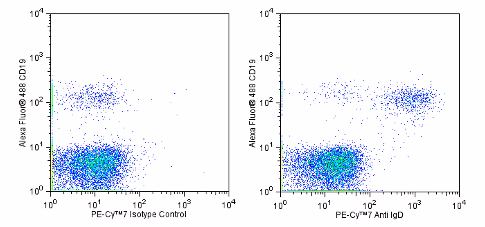
Flow cytometric analysis of IgD expression on human peripheral blood lymphocytes. Human peripheral blood mononuclear cells were cultured in complete tissue culture medium overnight in order to minimize subsequent nonspecific immunofluorescent staining. The cells were harvested and stained with Alexa Fluor® 488 Mouse anti-Human CD19 antibody (Cat. No. 557697) and with either a PE-Cy™7 Mouse IgG2a, κ Isotype Control (Cat. No. 552868; Left Panel) or a PE-Cy™7 Mouse anti-Human IgD antibody (Cat. No. 561314; Right Panel). The two-color flow cytometric dot plots showing the correlated expression of IgD (or Ig isotype control staining) versus CD19 were derived from events with the forward and side light-scatter characteristics of viable lymphocytes. Flow cytometry was performed using a BD™ LSR II Flow Cytometer System.
Flow cytometric analysis of IgD expression on human peripheral blood lymphocytes. Human peripheral blood mononuclear cells were cultured in complete tissue culture medium overnight in order to minimize subsequent nonspecific immunofluorescent staining. The cells were harvested and stained with Alexa Fluor® 488 Mouse anti-Human CD19 antibody (Cat. No. 557697) and with either a PE-Cy™7 Mouse IgG2a, κ Isotype Control (Cat. No. 552868; Left Panel) or a PE-Cy™7 Mouse anti-Human IgD antibody (Cat. No. 561314; Right Panel). The two-color flow cytometric dot plots showing the correlated expression of IgD (or Ig isotype control staining) versus CD19 were derived from events with the forward and side light-scatter characteristics of viable lymphocytes. Flow cytometry was performed using a BD™ LSR II Flow Cytometer System.


 全部商品分类


 下载产品说明书
 用小程序,查商品更便捷


 收藏
收藏
 对比
对比 咨询
咨询
