BD Horizon™ BV421 Goat Anti-Rabbit IgG is intended to be a second-step reagent for immunofluorescent staining of cells pre-stained with Rabbit IgG primary antibodies. It reacts with rabbit IgG and the light chains of other rabbit immunoglobulins. BV421 Anti-Rabbit IgG has minimal crossreactivity with human, mouse, or rat serum proteins. As a second step staining reagent, it is reactive with rabbit polyclonal and monoclonal antibodies.
The antibody was conjugated to BD Horizon BV421 which is part of the BD Horizon Brilliant™ Violet family of dyes. BD Horizon BV421 has an Ex Max at 407 nm and Em Max at 421 nm. The use of a mounting reagent (eg, ProLong® Gold) is highly recommended to maximize the photostability of BV421.
For confocal microscopy systems, a 405 nm laser is the optimal excitation source with optimal emission collection centered at 421 nm. For epifluorescence microscopes with broad spectrum excitation sources, the recommended excitation and emission filters are 392/23 nm and 430/24 nm bandpass filters, respectively. For specific multicolor imaging applications, the exact filter configurations should be optimized by the end user. For additional instrument/filter configuration information, please visit http://www.bdbiosciences.com/research/cellularimaging.
商品描述
Poly1272
BD Horizon™ BV421 Goat Anti-Rabbit IgG is intended to be a second-step reagent for immunofluorescent staining of cells pre-stained with Rabbit IgG primary antibodies. It reacts with rabbit IgG and the light chains of other rabbit immunoglobulins. BV421 Anti-Rabbit IgG has minimal crossreactivity with human, mouse, or rat serum proteins. As a second step staining reagent, it is reactive with rabbit polyclonal and monoclonal antibodies.
The antibody was conjugated to BD Horizon BV421 which is part of the BD Horizon Brilliant™ Violet family of dyes. BD Horizon BV421 has an Ex Max at 407 nm and Em Max at 421 nm. The use of a mounting reagent (eg, ProLong® Gold) is highly recommended to maximize the photostability of BV421.
For confocal microscopy systems, a 405 nm laser is the optimal excitation source with optimal emission collection centered at 421 nm. For epifluorescence microscopes with broad spectrum excitation sources, the recommended excitation and emission filters are 392/23 nm and 430/24 nm bandpass filters, respectively. For specific multicolor imaging applications, the exact filter configurations should be optimized by the end user. For additional instrument/filter configuration information, please visit http://www.bdbiosciences.com/research/cellularimaging.
同种型
Goat Ig
克隆号
克隆 Polyclonal (RUO)
浓度
0.2 mg/ml
产品详情
BV421
The BD Horizon Brilliant Violet™ 421 (BV421) Dye is part of the BD Horizon Brilliant Violet™ family of dyes. This polymer-technology based dye has an excitation maximum (Ex Max) of 407-nm and an emission maximum (Em Max) at 423-nm. Driven by BD innovation, BV421 is designed to be excited by the violet laser (405-nm) and detected using an optical filter centered near 420-nm (e.g., a 431/28-nm or 450/50-nm bandpass filter). BV421 is an ideal alternative for V450 as it is approximately ten times brighter with less spillover into the BV510/V500 detector. Please ensure that your instrument’s configurations (lasers and optical filters) are appropriate for this dye.
BV421
Violet 405 nm
407 nm
423 nm
应用
实验应用
Flow cytometry (Routinely Tested), Immunofluorescence (Tested During Development)
反应种属
Rabbit (QC Testing)
目标/特异性
IgG
制备和贮存
存储溶液
Aqueous buffered solution containing BSA and ≤0.09% sodium azide.
保存方式
Aqueous buffered solution containing BSA and ≤0.09% sodium azide.
参考图片
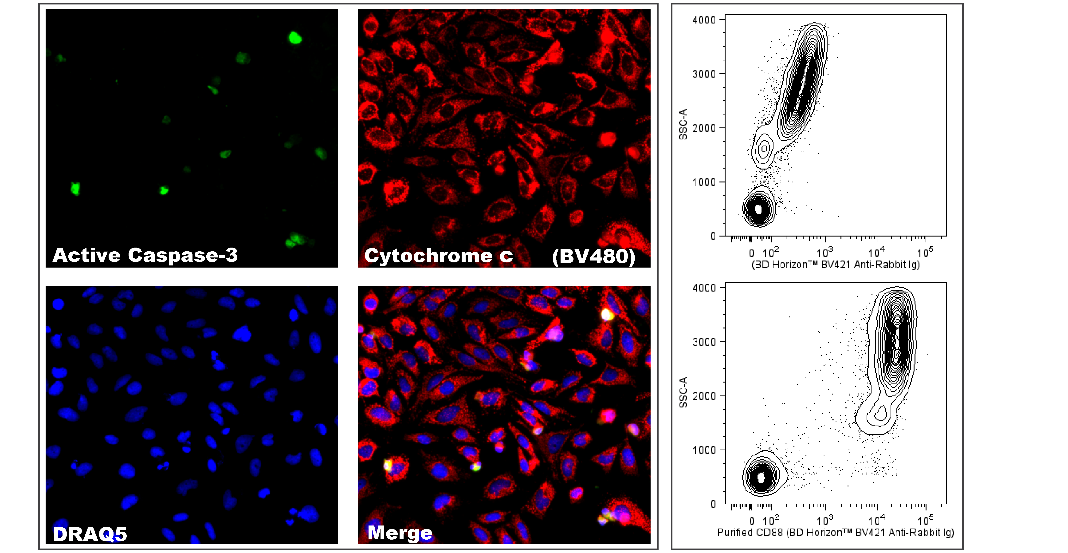
Immunofluorescent Staining using BD Horizon™ BV421 Goat Anti-Rabbit Ig Left Panel - Immunofluorescence staining of apoptotic cells. HeLa cells were treated with Camptothecin (20 µM, 6 hr) to induce apoptosis. Cells were fixed with BD Cytofix™ Fixation Buffer (Cat. No. 554655), permeabilized (5 min) with BD Phosflow™ Perm Buffer III (Cat. No. 558050), washed with 1× PBS, and blocked (30 min) with 5% Goat serum, 1% BSA, and 0.5% Triton™ X-100 diluted in 1× PBS. The cells were stained (1 hr) with Purified Mouse Anti-Cytochrome c (Cat. No. 556432) and Purified Rabbit Anti-Active Caspase-3 (Cat. No. 559565) antibodies at 2.5 µg/mL diluted in blocking buffer. After washing, the cells were stained (45 min) with the second step reagents, BD Horizon™ BV421 Goat Anti-Rabbit Ig (Cat. No. 565014; pseudo-colored green) and BD Horizon™ BV480 Goat Anti-Mouse Ig (Cat. No. 564877, pseudo-colored red) antibodies at 2.5 µg/mL diluted in blocking buffer. DRAQ5 (Cat. No. 564902/564903; pseudo-colored blue) was used as a nuclear counterstain. Images were acquired with a standard epifluorescence microscope with a 20× objective. Right Panel - Multiparameter flow cytometric analysis of CD88 expression on human peripheral blood leucocytes. Whole blood was either not stained with a primary antibody as a control (Top Plot) or primarily stained with Purified Rabbit Anti-Human CD88 monoclonal antibody (Cat. No. 559159; Bottom Plot). Erythrocytes were lysed with BD FACS Lysing Solution (Cat. No. 349202). The cells were washed and secondarily stained with BD Horizon™ BV421 Goat Anti-Rabbit Ig (Cat. No. 565014). Two-parameter flow cytometric contour plots showing the correlated expression of CD88 (or Unstained control) versus side light-scatter (SSC-A) signals were derived from gated events with the forward and side light-scatter characteristics of intact leucocyte populations. Flow cytometric analysis was performed using a BD LSRFortessa™ Cell Analyzer System.


 全部商品分类


 下载产品说明书
 用小程序,查商品更便捷


 收藏
收藏
 对比
对比 咨询
咨询
