


 全部商品分类


 用小程序,查商品更便捷


Monoclonal antibody is produced by immunizing animals with recombinant human IL-1β protein.

Product Usage Information
| Application | Dilution |
|---|---|
| Western Blotting | 1:1000 |
| Simple Western™ | 1:10 - 1:50 |
| Immunofluorescence (Immunocytochemistry) | 1:200 - 1:800 |
| Flow Cytometry (Fixed/Permeabilized) | 1:100 - 1:400 |
Specificity/Sensitivity
Species Reactivity:
Human
Supplied in 10 mM sodium HEPES (pH 7.5), 150 mM NaCl, 100 µg/ml BSA, 50% glycerol and less than 0.02% sodium azide. Store at –20°C. Do not aliquot the antibody.
For a carrier free (BSA and azide free) version of this product see product #18689.
参考图片
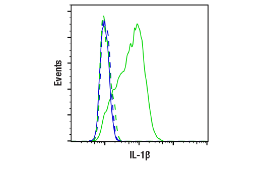
Flow cytometric analysis of THP-1, untreated (blue, negative) or treated with LPS #14011 (100 ng/ml, 3 hr; green, positive) using IL-1β (D3U3E) Rabbit mAb (solid lines) or concentration-matched Rabbit (DA1E) mAb IgG XP® Isotype Control #3900 (dashed lines). Anti-rabbit IgG (H+L), F(ab')₂ Fragment (Alexa Fluor® 488 Conjugate) #4412 was used as a secondary antibody.
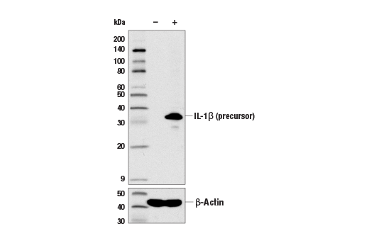
Western blot analysis of extracts from THP-1 cells, untreated (-) or LPS-treated (100 ng/ml, 3 hr; +), using IL-1β (D3U3E) Rabbit mAb (upper) and β-Actin (D6A8) Rabbit mAb #8457 (lower).
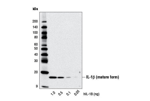
Western blot analysis of recombinant Human Interleukin-1β (hIL-1β) #8900 using IL-1β (D3U3E) Rabbit mAb.
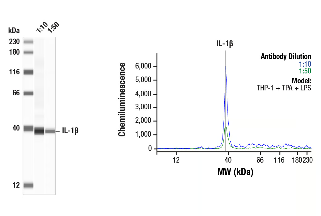
Simple Western™ analysis of lysates (0.1 mg/mL) from THP-1 cells treated with TPA (80nM, O/N) + LPS (1 µg/ml 15min) using IL-1β (D3U3E) Rabbit mAb #12703. The virtual lane view (left) shows the target band (as indicated) at 1:10 and 1:50 dilutions of primary antibody. The corresponding electropherogram view (right) plots chemiluminescence by molecular weight along the capillary at 1:10 (blue line) and 1:50 (green line) dilutions of primary antibody. This experiment was performed under reducing conditions on the Jess™ Simple Western instrument from ProteinSimple, a BioTechne brand, using the 12-230 kDa separation module.
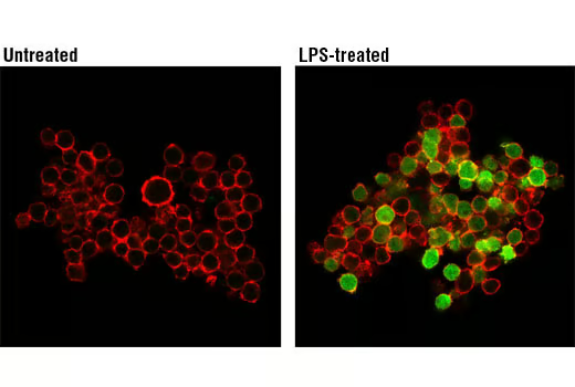
Confocal immunofluorescent analysis of THP-1 cells, untreated (left) or LPS-treated (500 ng/ml, 2 hr; right), using IL-1β (D3U3E) Rabbit mAb (green). Actin filaments were labeled with DY-554 phalloidin (red).