Human IL-17A, also known as IL-17, is a proinflammatory cytokine that is encoded by the IL17A gene in chromosome 6. IL-17A is produced as a disulfide-linked homodimer comprised of two mature 136-amino acid polypeptides. It is a member of the IL-17 family of structurally related cytokines, designated IL-17A through IL-17F. Activated memory T cells, especially Th17 cells (specialized IL-17A-producing CD4+ T cells distinct from Th1 and Th2 cells) produce IL-17 and provide protective immunity against pathogens. Activated CD8+ T cells, γδT cells, NK cells and neutrophils can also be activated to produce IL-17A. IL-17A binds to and exerts its biological activity through IL-17 receptors (IL-17R) that are expressed by a variety of target cells including fibroblasts, epithelial and endothelial cells, monocytes/macrophages and mast cells. The ubiquitous IL-17R expression pattern may explain the broad tissue responsiveness to IL-17. IL-17 induces stromal cells to secrete cytokines and chemokines involved in inflammatory and hematopoietic processes. For example, IL-17 induces fibroblasts to produce IL-6, IL-8, G-CSF and express increased surface ICAM-1. The N49-653 antibody reacts with human IL-17A.
The antibody was conjugated to BD Horizon™ BV421 which is part of the BD Horizon™ Brilliant Violet™ family of dyes. With an Ex Max of 407-nm and Em Max at 421-nm, BD Horizon™ BV421 can be excited by the violet laser and detected in the standard Pacific Blue™ filter set (eg, 450/50-nm filter). BD Horizon™ BV421 conjugates are very bright, often exhibiting a 10 fold improvement in brightness compared to Pacific Blue™ conjugates.
商品描述
N49-653
Human IL-17A, also known as IL-17, is a proinflammatory cytokine that is encoded by the IL17A gene in chromosome 6. IL-17A is produced as a disulfide-linked homodimer comprised of two mature 136-amino acid polypeptides. It is a member of the IL-17 family of structurally related cytokines, designated IL-17A through IL-17F. Activated memory T cells, especially Th17 cells (specialized IL-17A-producing CD4+ T cells distinct from Th1 and Th2 cells) produce IL-17 and provide protective immunity against pathogens. Activated CD8+ T cells, γδT cells, NK cells and neutrophils can also be activated to produce IL-17A. IL-17A binds to and exerts its biological activity through IL-17 receptors (IL-17R) that are expressed by a variety of target cells including fibroblasts, epithelial and endothelial cells, monocytes/macrophages and mast cells. The ubiquitous IL-17R expression pattern may explain the broad tissue responsiveness to IL-17. IL-17 induces stromal cells to secrete cytokines and chemokines involved in inflammatory and hematopoietic processes. For example, IL-17 induces fibroblasts to produce IL-6, IL-8, G-CSF and express increased surface ICAM-1. The N49-653 antibody reacts with human IL-17A.
The antibody was conjugated to BD Horizon™ BV421 which is part of the BD Horizon™ Brilliant Violet™ family of dyes. With an Ex Max of 407-nm and Em Max at 421-nm, BD Horizon™ BV421 can be excited by the violet laser and detected in the standard Pacific Blue™ filter set (eg, 450/50-nm filter). BD Horizon™ BV421 conjugates are very bright, often exhibiting a 10 fold improvement in brightness compared to Pacific Blue™ conjugates.
同种型
Mouse IgG1, κ
克隆号
克隆 N49-653 (RUO)
产品详情
BV421
The BD Horizon Brilliant Violet™ 421 (BV421) Dye is part of the BD Horizon Brilliant Violet™ family of dyes. This polymer-technology based dye has an excitation maximum (Ex Max) of 407-nm and an emission maximum (Em Max) at 423-nm. Driven by BD innovation, BV421 is designed to be excited by the violet laser (405-nm) and detected using an optical filter centered near 420-nm (e.g., a 431/28-nm or 450/50-nm bandpass filter). BV421 is an ideal alternative for V450 as it is approximately ten times brighter with less spillover into the BV510/V500 detector. Please ensure that your instrument’s configurations (lasers and optical filters) are appropriate for this dye.
Aqueous buffered solution containing BSA and ≤0.09% sodium azide.
保存方式
Aqueous buffered solution containing BSA and ≤0.09% sodium azide.
文献
文献
研发参考(6)
1. Fossiez F, Djossou O, Chomarat P, et al. T cell interleukin-17 induces stromal cells to produce proinflammatory and hematopoietic cytokines. J Exp Med. 1996; 183(6):2593-2603. (Biology).
2. Korn T, Oukka M, Kuchroo V, Bettelli E. Th17 cells: effector T cells with inflammatory properties. Semin Immunol. 2007; 19(6):362-371. (Biology).
3. Moseley TA, Haudenschild DR, Rose L, Reddi AH. Interleukin-17 family and IL-17 receptors. Cytokine Growth Factor Rev. 2003; 14(2):155-174. (Biology).
4. Weaver CT, Hatton RD, Mangan PR, Harrington LE. IL-17 family cytokines and the expanding diversity of effector T cell lineages. Annu Rev Immunol. 2007; 25:821-852. (Biology).
5. Yao Z, Painter SL, Fanslow WC, et al. Human IL-17: a novel cytokine derived from T cells. J Immunol. 1995; 155(12):5483-5486. (Biology).
6. Yao Z, Spriggs MK, Derry JM, et al. Molecular characterization of the human interleukin (IL)-17 receptor. Cytokine. 1997; 9(11):794-800. (Biology).
参考图片
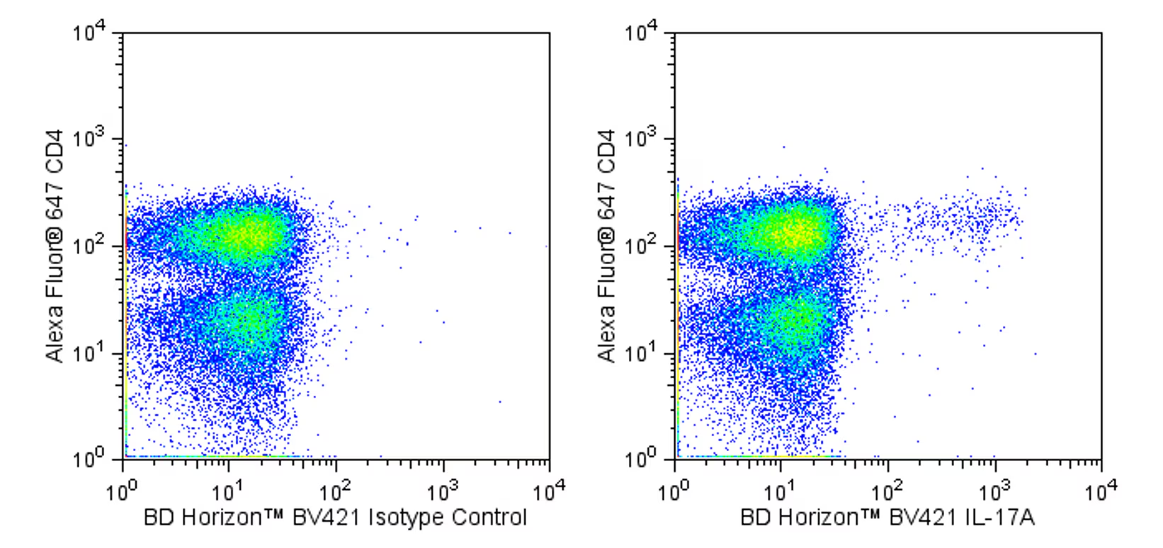
Multicolor flow cytometric analysis of IL-17A expressed in stimulated human peripheral blood mononuclear cells. HiCK-1 Human Cytokine Positive Control Cells (Cat. No. 555061) were permeabilized with BD Perm/Wash™ Buffer (Cat. No. 554723). The cells were then stained with either a BD Horizon™ BV421 Mouse IgG1, κ Isotype Control (Cat No. 562438, Left Panel) or with the BD Horizon™ BV421 Mouse Anti-Human IL-17A antibody (Cat No. 562933, Right Panel) in conjunction with an Alexa Fluor® 647 Mouse Anti-Human CD4 antibody (Cat. No. 557707). Two-color flow cytometric dot plots showing the expression of IL-17A (or Ig Isotype Control staining) versus CD4 were derived from gated events with the forward and side light-scatter characteristics of intact lymphocytes. Flow cytometry was performed using a BD LSR™ II Flow Cytometry System.
Multicolor flow cytometric analysis of IL-17A expressed in stimulated human peripheral blood mononuclear cells. HiCK-1 Human Cytokine Positive Control Cells (Cat. No. 555061) were permeabilized with BD Perm/Wash™ Buffer (Cat. No. 554723). The cells were then stained with either a BD Horizon™ BV421 Mouse IgG1, κ Isotype Control (Cat No. 562438, Left Panel) or with the BD Horizon™ BV421 Mouse Anti-Human IL-17A antibody (Cat No. 562933, Right Panel) in conjunction with an Alexa Fluor® 647 Mouse Anti-Human CD4 antibody (Cat. No. 557707). Two-color flow cytometric dot plots showing the expression of IL-17A (or Ig Isotype Control staining) versus CD4 were derived from gated events with the forward and side light-scatter characteristics of intact lymphocytes. Flow cytometry was performed using a BD LSR™ II Flow Cytometry System.


 全部商品分类


 下载产品说明书
 用小程序,查商品更便捷


 收藏
收藏
 对比
对比 咨询
咨询
