The TC11-18H10 monoclonal antibody specifically binds to recombinant and natural mouse IL-17A proteins. IL-17A, also known as CTLA-8, is a T cell-derived cytokine that promotes inflammatory responses. Mouse IL-17A is a proinflammatory cytokine that can induce the release of IL-6 by mouse stromal cells. It has been shown to support the growth of hemopoietic progenitors in vitro; it can also stimulate granulopoiesis in vivo. The TC11-18H10 antibody has been reported to neutralize IL-17A activity. Recent studies have shown that IL-17A is produced by a unique subset of Th17 cells that develop along a pathway distinct from the Th1- and Th2- cell differentiation pathways. The mouse IL-17A cDNA was isolated from a cDNA library generated from TCRαβ+CD4-CD8- thymocytes.
商品描述
TC11-18H10
The TC11-18H10 monoclonal antibody specifically binds to recombinant and natural mouse IL-17A proteins. IL-17A, also known as CTLA-8, is a T cell-derived cytokine that promotes inflammatory responses. Mouse IL-17A is a proinflammatory cytokine that can induce the release of IL-6 by mouse stromal cells. It has been shown to support the growth of hemopoietic progenitors in vitro; it can also stimulate granulopoiesis in vivo. The TC11-18H10 antibody has been reported to neutralize IL-17A activity. Recent studies have shown that IL-17A is produced by a unique subset of Th17 cells that develop along a pathway distinct from the Th1- and Th2- cell differentiation pathways. The mouse IL-17A cDNA was isolated from a cDNA library generated from TCRαβ+CD4-CD8- thymocytes.
同种型
Rat LEW, also known as Lewis IgG1, κ
克隆号
克隆 TC11-18H10 (RUO)
浓度
0.2 mg/ml
产品详情
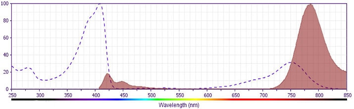
BV786
The BD Horizon Brilliant Violet™ 786 (BV786) Dye is part of the BD Horizon Brilliant Violet™ family of dyes. This tandem fluorochrome is comprised of a BV421 donor with an Ex Max of 407-nm and an acceptor dye with an Em Max at 786-nm. BV786, driven by BD innovation, is designed to be excited by the violet laser and detected using a filter, centered near 785 nm (e.g. 780/60 nm bandpass filter). Please ensure that your instrument’s configurations (lasers and filters) are appropriate for this dye.
Aqueous buffered solution containing BSA and ≤0.09% sodium azide.
保存方式
Aqueous buffered solution containing BSA and ≤0.09% sodium azide.
文献
文献
研发参考(4)
1. Coquet JM, Chakravarti S, Kyparissoudis K, et al. Diverse cytokine production by NKT cell subsets and identification of an IL-17-producing CD4-NK1.1- NKT cell population. Proc Natl Acad Sci U S A. 2008; 105(32):11287-11292. (Clone-specific: Flow cytometry).
2. Dong C. Th17 cells: Current understanding of their generation and regulation. Eur J Immunol. 2009; 39(3):640-644. (Biology).
3. Prussin C, Metcalfe DD. Detection of intracytoplasmic cytokine using flow cytometry and directly conjugated anti-cytokine antibodies. J Immunol Methods. 1995; 188(1):117-128. (Methodology: Flow cytometry).
4. Schwarzenberger P, La Russa V, Miller A, et al. IL-17 stimulates granulopoiesis in mice: use of an alternate, novel gene therapy-derived method for in vivo evaluation of cytokines. J Immunol. 1998; 161(11):6383-6389. (Biology).
参考图片
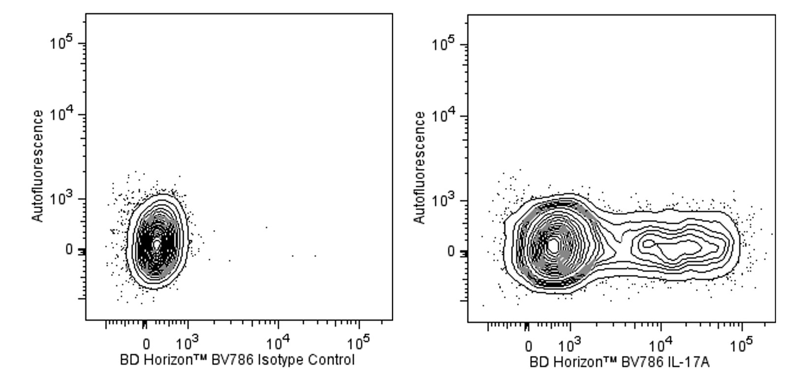
Flow cytometric analysis of IL-17A-producing mouse EL-4 cells. Cells from the mouse EL4 (T lymphoma, ATCC TIB-39) cell line were stimulated (5 hr) with Phorbol 12-Myristate 13-Acetate (PMA; 50 ng/ml final concentration; Sigma, Cat. No. P-8139) and Ionomycin (1000 ng/ml final concentration; Sigma, Cat. No. I-0634) in the presence of BD GolgiStop™ Protein Transport Inhibitor (containing Monensin) (Cat. No. 554724). The cells were washed with BD Pharmingen™ Stain Buffer (FBS) (Cat. No. 554656), and fixed and permeabilized with BD Cytofix/Cytoperm™ Fixation and Permeabilization Solution (Cat. No. 554722).
The cells were then washed and stained in BD Perm/Wash™ Buffer (Cat. No. 554723) with BD Horizon™ BV786 Rat IgG1 κ Isotype Control (Cat. No. 563847; Left Panel) or BD Horizon BV786 Rat Anti-Mouse IL-17A antibody (Cat. No. 564171; Right Panel) using BD Biosciences' Protocol for Immunofluorescent Staining of Intracellular Cytokines for Flow Cytometric Analysis. Two-color contour plots showing the expression of IL-17A (or Ig Isotype control staining) versus Autofluorescence were derived from gated events with the forward and side light-scatter characteristics of intact cells. Flow cytometric analysis was performed using a BD™ LSR II Flow Cytometer System.
Flow cytometric analysis of IL-17A-producing mouse EL-4 cells. Cells from the mouse EL4 (T lymphoma, ATCC TIB-39) cell line were stimulated (5 hr) with Phorbol 12-Myristate 13-Acetate (PMA; 50 ng/ml final concentration; Sigma, Cat. No. P-8139) and Ionomycin (1000 ng/ml final concentration; Sigma, Cat. No. I-0634) in the presence of BD GolgiStop™ Protein Transport Inhibitor (containing Monensin) (Cat. No. 554724). The cells were washed with BD Pharmingen™ Stain Buffer (FBS) (Cat. No. 554656), and fixed and permeabilized with BD Cytofix/Cytoperm™ Fixation and Permeabilization Solution (Cat. No. 554722). The cells were then washed and stained in BD Perm/Wash™ Buffer (Cat. No. 554723) with BD Horizon™ BV786 Rat IgG1 κ Isotype Control (Cat. No. 563847; Left Panel) or BD Horizon BV786 Rat Anti-Mouse IL-17A antibody (Cat. No. 564171; Right Panel) using BD Biosciences' Protocol for Immunofluorescent Staining of Intracellular Cytokines for Flow Cytometric Analysis. Two-color contour plots showing the expression of IL-17A (or Ig Isotype control staining) versus Autofluorescence were derived from gated events with the forward and side light-scatter characteristics of intact cells. Flow cytometric analysis was performed using a BD™ LSR II Flow Cytometer System.


 全部商品分类


 下载产品说明书
 用小程序,查商品更便捷


 收藏
收藏
 对比
对比 咨询
咨询