


 全部商品分类
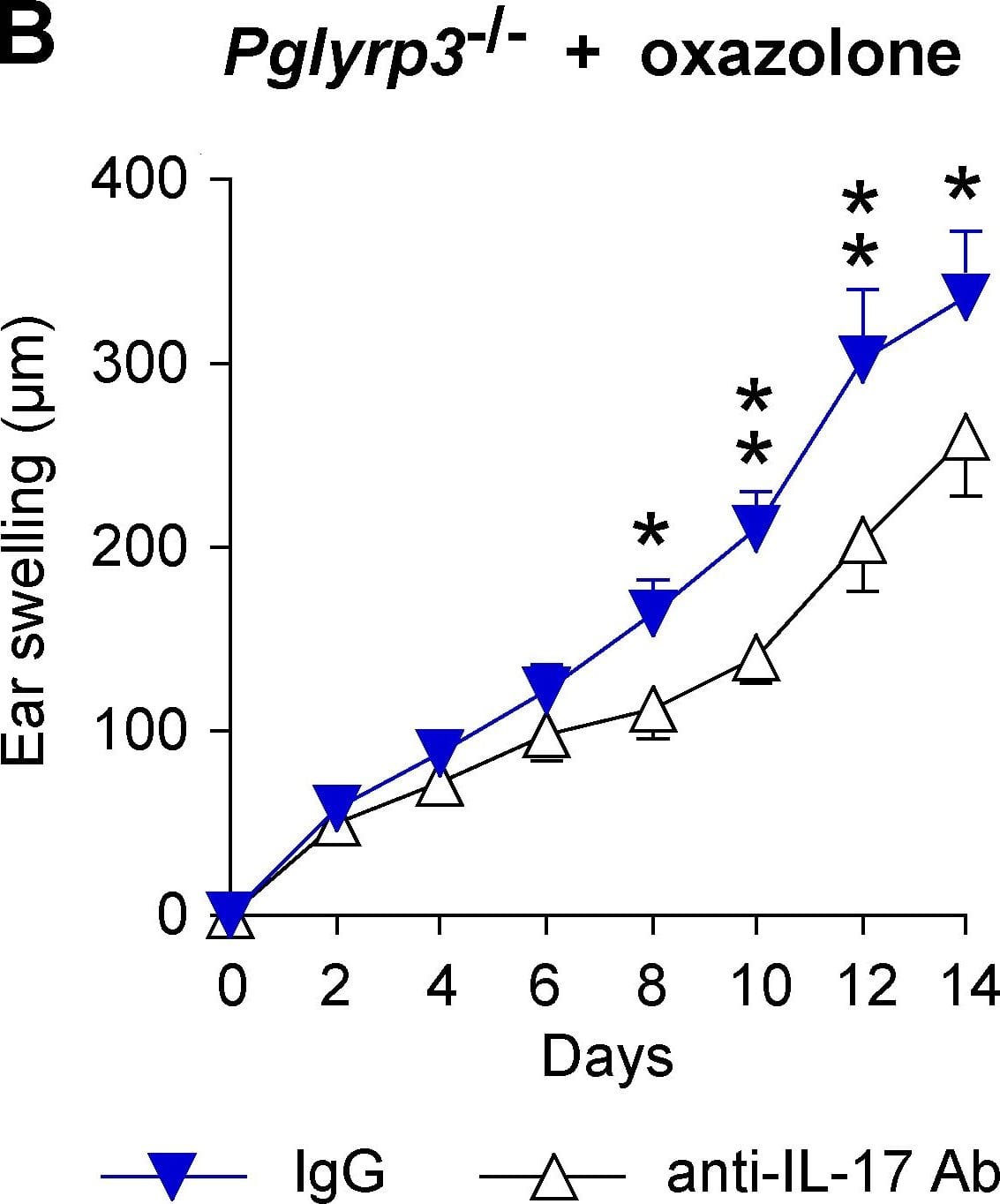


mIL-17 MAb (Cl 50104 (25 ug)


 下载产品说明书 下载SDS
 用小程序,查商品更便捷


 收藏
收藏
 对比
对比 咨询
咨询
Thr22-Ala158
Accession # Q62386
Scientific Data
 View Larger
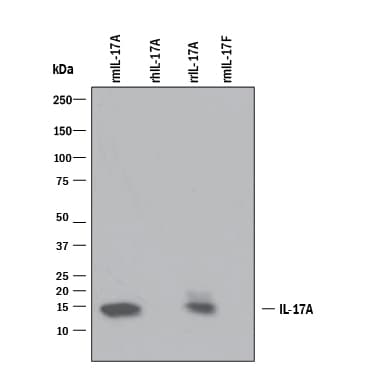
Detection of Recombinant Mouse IL‑17/IL‑17A by Western Blot. Western blot shows 25 ng of Recombinant Mouse IL-17/ IL-17A (Catalog # 421-ML), Recombinant Human IL-17/IL-17A (Catalog # 317-ILB), Recombinant Rat IL-17/IL-17A (Catalog # 8410-IL), and Recombinant Mouse IL-17F (Catalog # 2057-IL). PVDF Membrane was probed with 1 µg/mL of Rat Anti-Mouse IL-17/ IL-17A Monoclonal Antibody (Catalog # MAB421) followed by HRP-conjugated Anti-Rat IgG Secondary Antibody (Catalog # HAF005). A specific band was detected for IL-17/IL-17A at approximately 15 kDa (as indicated). This experiment was conducted under reducing conditions and using Immunoblot Buffer Group 3.
 View Larger
IL‑6 Secretion Induced by IL‑17 and Neutralization by Mouse IL‑17 Antibody. Recombinant Mouse IL-17 (Catalog # 421-ML) stimulates IL-6 secretion in the NIH-3T3 mouse embryonic fibroblast cell line in a dose-dependent manner (orange line), as measured by the Mouse IL-6 Quantikine ELISA Kit (Catalog # M6000B). IL-6 secretion elicited by Recombinant Mouse IL-17 (10 ng/mL) is neutralized (green line) by increasing concentrations of Rat Anti-Mouse IL-17 Monoclonal Antibody (Catalog # MAB421). The ND50 is typically 0.05-0.15 µg/mL.
 View Larger
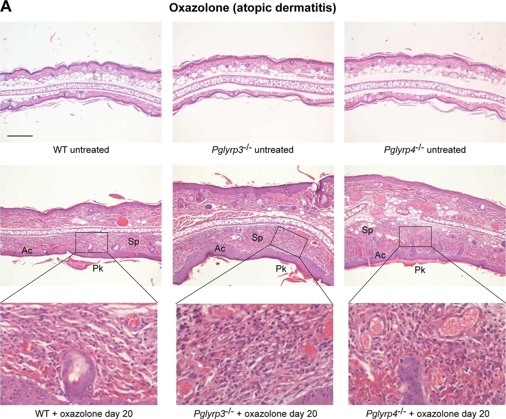
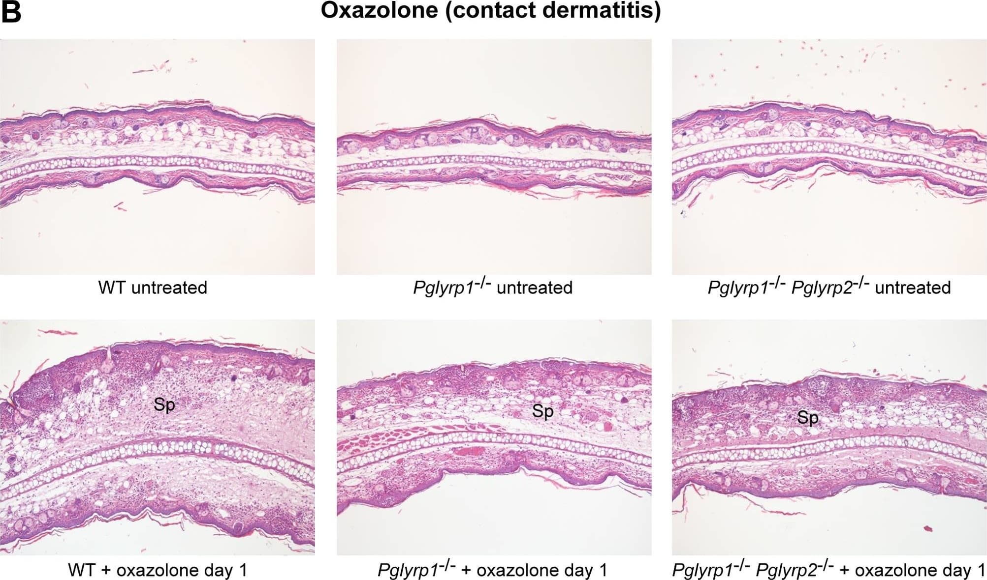
Detection of Mouse IL-17/IL-17A by In vivo assay Ear histology in WT and Pglyrp-deficient mice in atopic dermatitis and contact dermatitis models of skin inflammation.(A) Oxazolone model of atopic dermatitis: sensitization and 10 applications of oxazolone to the ears every other day induced acanthosis (Ac), parakeratosis (Pk), marked thickening of the sub-epidermal layer with spongiosis (Sp) and dense cellular infiltrates of primarily mononuclear and some polymorphonuclear cells (high magnification insets), that were all highly prominent in Pglyrp3−/− mice and Pglyrp4−/− mice and much less severe in WT mice. (B) Oxazolone model of contact dermatitis: sensitization and a single application of oxazolone to the ears induced strong inflammatory response in WT mice with marked spongiosis of the sub-epidermal layer (Sp) and cellular infiltrates of epidermal and sub-epidermal layers, composed of mononuclear and polymorphonuclear cells; Pglyrp1−/− and Pglyrp1−/−Pglyrp2−/− mice still had cellular infiltrates, but had substantially reduced swelling, compared to WT mice, mostly due to reduced edema. H&E stained cross-sections; bar = 200 µm for all panels, except high magnification insets (the magnified areas are shown by rectangles). Image collected and cropped by CiteAb from the following publication (https://pubmed.ncbi.nlm.nih.gov/21949809), licensed under a CC-BY license. Not internally tested by R&D Systems.
 View Larger
Detection of Mouse IL-17/IL-17A by In vivo assay IL-17 is required for enhanced response to oxazolone in Pglyrp3−/− mice.(A) The level of IL-17-induced chemokine, CXCL-1, is higher in the ears of Pglyrp3−/− mice than WT mice after sensitization and application of oxazolone for 20 days. (B) Ear swelling in Pglyrp3−/− mice sensitized and treated 7 times with oxazolone every other day and also treated with neutralizing anti-IL-17 mAbs is lower than in Pglyrp3−/−mice similarly treated with oxazolone and isotype control IgG. Means ± SEM; N = 6 mice/group; significance of differences between Pglyrp3−/− and WT mice (A) or IgG control and anti-IL-17 mAbs-treated mice (B): *, P<0.05; **, P<0.005. Image collected and cropped by CiteAb from the following publication (https://pubmed.ncbi.nlm.nih.gov/21949809), licensed under a CC-BY license. Not internally tested by R&D Systems.
 View Larger
Detection of Mouse IL-17/IL-17A by In vivo assay Ear histology in WT and Pglyrp-deficient mice in atopic dermatitis and contact dermatitis models of skin inflammation.(A) Oxazolone model of atopic dermatitis: sensitization and 10 applications of oxazolone to the ears every other day induced acanthosis (Ac), parakeratosis (Pk), marked thickening of the sub-epidermal layer with spongiosis (Sp) and dense cellular infiltrates of primarily mononuclear and some polymorphonuclear cells (high magnification insets), that were all highly prominent in Pglyrp3−/− mice and Pglyrp4−/− mice and much less severe in WT mice. (B) Oxazolone model of contact dermatitis: sensitization and a single application of oxazolone to the ears induced strong inflammatory response in WT mice with marked spongiosis of the sub-epidermal layer (Sp) and cellular infiltrates of epidermal and sub-epidermal layers, composed of mononuclear and polymorphonuclear cells; Pglyrp1−/− and Pglyrp1−/−Pglyrp2−/− mice still had cellular infiltrates, but had substantially reduced swelling, compared to WT mice, mostly due to reduced edema. H&E stained cross-sections; bar = 200 µm for all panels, except high magnification insets (the magnified areas are shown by rectangles). Image collected and cropped by CiteAb from the following publication (https://pubmed.ncbi.nlm.nih.gov/21949809), licensed under a CC-BY license. Not internally tested by R&D Systems.
Mouse IL-17/IL-17A Antibody Summary
(rm) IL-17A/IL-17F heterodimer is observed. No cross-reactivity with recombinant human IL-17, recombinant canine IL-17, rmIL-17B,
rmIL‑17C, rmIL-17D, rmIL-17E, or rmIL-17F is observed.
Thr22-Ala158
Accession # Q62386
Applications
Please Note: Optimal dilutions should be determined by each laboratory for each application. General Protocols are available in the Technical Information section on our website.
Background: IL-17/IL-17A
Interleukin 17 (also known as CTLA-8) is a T cell-expressed pleiotropic cytokine that exhibits a high degree of homology to a protein encoded by the ORF13 gene of herpes virus Saimiri. cDNA clones encoding IL-17 have been isolated from activated rat, mouse and human T cells. Mouse IL-17 cDNA encodes a 158 amino acid (aa) residue precursor protein with a 21 amino acid residue signal peptide that is cleaved to yield the 137 aa residue mature IL-17. Both recombinant and natural IL-17 have been shown to exist as disulfide linked homodimers. At the amino acid level, mIL-17 shows 57% and 87% sequence identity with herpesvirus and rat IL-17, respectively. An IL-17 specific mouse cell surface receptor (IL-17 R) has been cloned. While the expression of IL-17 mRNA is restricted to activated alpha beta TCR+CD4-CD8-T cells, the expression of mIL-17 R mRNA has been detected in virtually all cells and tissues tested. IL-17 exhibits multiple biological activities on a variety of cells including: the induction of IL-6 and IL-8 production in fibroblasts; the enhancement of surface expression of ICAM-1 in fibroblasts; activation of NF-kappa B and costimulation of T cell proliferation.
- Kennedy, J. et al. (1996) J. Interferon Cytokine Res. 16:611.
- Yao, Z. et al. (1995) J. Immunol. 155:5483.
- Yao, Z. et al. (1995) Immunity 3:811.
- Rouvier, E. et al. (1993) J. Immunol. 150:5445.
Preparation and Storage
- 12 months from date of receipt, -20 to -70 °C as supplied.
- 1 month, 2 to 8 °C under sterile conditions after reconstitution.
- 6 months, -20 to -70 °C under sterile conditions after reconstitution.
参考图片
IL‑6 Secretion Induced by
IL‑17 and Neutralization by Mouse IL‑17 Antibody. Recombinant Mouse IL‑17 (Catalog # 421-ML) stimulates IL‑6 secretion in the NIH‑3T3 mouse embryonic fibroblast cell line in a dose-dependent manner (orange line), as measured by the Mouse IL‑6 Quantikine ELISA Kit (Catalog # M6000B). IL‑6 secretion elicited by Recombinant Mouse IL‑17 (10 ng/mL) is neutralized (green line) by increasing concentrations of Rat Anti-Mouse IL‑17 Monoclonal Antibody (Catalog # MAB421). The ND50 is typically
0.05-0.15 µg/mL.