


 全部商品分类


IL-1beta (3A6) Mouse mAb

 下载产品说明书 下载COA 下载SDS
 用小程序,查商品更便捷


 收藏
收藏
 对比
对比 咨询
咨询Monoclonal antibody is produced by immunizing animals with recombinant human IL-1β protein.

Product Usage Information
| Application | Dilution |
|---|---|
| Western Blotting | 1:1000 |
| Simple Western™ | 1:10 - 1:50 |
| Immunohistochemistry (Paraffin) | 1:50 - 1:200 |
Specificity/Sensitivity
Species Reactivity:
Human, Mouse
Supplied in 10 mM sodium HEPES (pH 7.5), 150 mM NaCl, 100 µg/ml BSA, 50% glycerol and less than 0.02% sodium azide. Store at –20°C. Do not aliquot the antibody.
For a carrier free (BSA and azide free) version of this product see product #27989.
参考图片
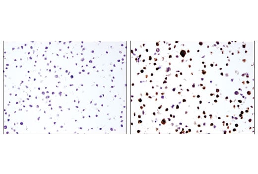
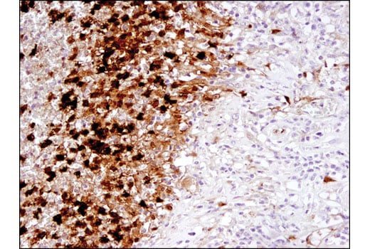
Immunohistochemical analysis of paraffin embedded THP-1 cell pellets, control (left) or LPS-treated (right) using IL-1-β (3A6) Mouse mAb.
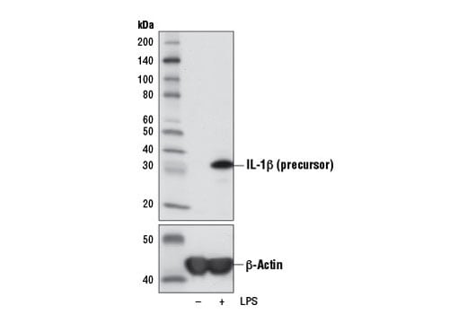
Western blot analysis of extracts from THP-1 cells, untreated (-) or LPS-treated (100 ng/mL, 3 hr; +), using IL-1β (3A6) Mouse mAb (upper) or β-Actin (D6A8) Rabbit mAb #8457 (lower).
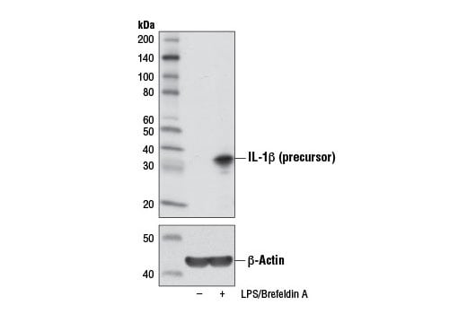
Western blot analysis of extracts from Raw 264.7 cells, untreated (-) or treated with Brefeldin A #9972 (300 ng/mL, last 3 hr of stimulation; +) and LPS (100 ng/mL, 6 hr; +), using IL-1β (3A6) Mouse mAb (upper) or β-Actin (D6A8) Rabbit mAb #8457 (lower).
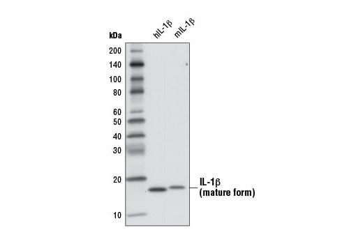
Western blot analysis of 1 ng recombinant Human Interleukin-1β (hIL-1β) #8900 and 1 ng recombinant Mouse Interleukin-1β (mIL-1β) #5204 using IL-1β (3A6) Mouse mAb.
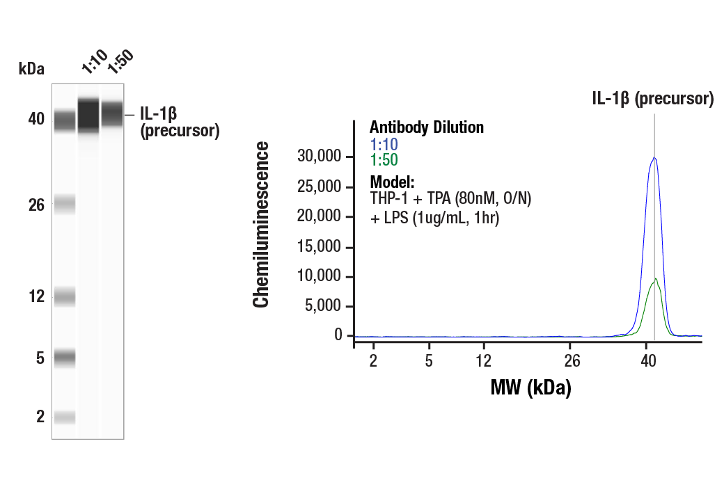
Simple Western™ analysis of lysates (1.0 mg/mL) from THP-1 cells treated with TPA (80nM, O/N) and LPS (1ug/ml, 1 hr) using IL-1β (3A6) Mouse mAb #12242. The virtual lane view (left) shows the target band (as indicated) at 1:10 and 1:50 dilutions of primary antibody. The corresponding electropherogram view (right) plots chemiluminescence by molecular weight along the capillary at 1:10 (blue line) and 1:50 (green line) dilutions of primary antibody. This experiment was performed under reducing conditions on the Jess™ Simple Western instrument from ProteinSimple, a BioTechne brand, using the 2-40 kDa separation module.
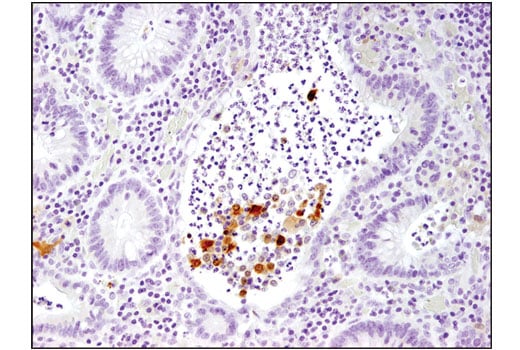
Immunohistochemical analysis of paraffin-embedded human large intestine (chronic colitis of the colon) using IL-1-β (3A6) Mouse mAb.
Immunohistochemical analysis of paraffin-embedded human large intestine (ulcerative chronic colitis of the rectum) using IL-1-β (3A6) Mouse mAb.