


 全部商品分类


hIL-1b QKit (1 Kit)


 下载产品说明书 下载SDS
 用小程序,查商品更便捷


 收藏
收藏
 对比
对比 咨询
咨询Sample Values
Serum/Plasma - Forty serum and plasma samples from apparently healthy volunteers were evaluated for the presence of human IL-1 beta in this assay. No medical histories were available for the donors used in this study. All samples measured less than the lowest IL-1 beta standard, 3.9 pg/mL.| Stimulant | Day 1 (pg/mL) | Day 3 (pg/mL) | Day 5 (pg/mL) |
| 10 μg/mL PHA | 2185 | 2004 | 2383 |
| 10 μg/mL PHA+10 ng/mL rhIL-2 | 1938 | 1973 | 2839 |
| 50 ng/mL PMA | 1767 | 1027 | 1159 |
| 50 ng/mL LPS | 4158 | 2145 | 1308 |
Recovery
The recovery of IL-1 beta spiked to different levels throughout the range of the assay in various matrices was evaluated.
| Sample Type | Average % Recovery | Range % |
|---|---|---|
| Cell Culture Media (n=4) | 97 | 80-111 |
| Citrate Plasma (n=10) | 93 | 83-110 |
| EDTA Plasma (n=10) | 86 | 81-100 |
| Heparin Plasma (n=10) | 82 | 76-100 |
| Serum (n=10) | 95 | 87-110 |
Linearity

Scientific Data


Assay Procedure
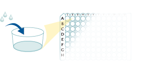
Refer to the product- Prepare all reagents, standard dilutions, and samples as directed in the product insert.
- Remove excess microplate strips from the plate frame, return them to the foil pouch containing the desiccant pack, and reseal.
- For Serum & Plasma Samples: Add 50 µL of Assay Diluent to each well.
- Add 200 µL of Standard, control, or sample to each well. Cover with a plate sealer, and incubate at room temperature for 2 hours.
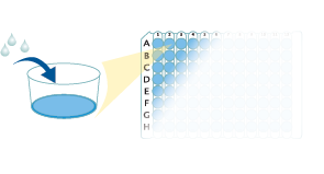
- Aspirate each well and wash, repeating the process twice for a total of 3 washes.
- Add 200 µL of Conjugate to each well.
- For Cell Culture Supernate Samples: Cover with a new plate sealer, and incubate at room temperature for 1 hour.
For Serum & Plasma Samples: Cover with a new plate sealer, and incubate at room temperature for 2 hours. - Aspirate and wash 3 times.
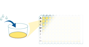
- Add 200 µL Substrate Solution to each well. Incubate at room temperature for 20 minutes. PROTECT FROM LIGHT.
- Add 50 µL of Stop Solution to each well. Read at 450 nm within 30 minutes. Set wavelength correction to 540 nm or 570 nm.
Human IL-1 beta/IL-1F2 Quantikine ELISA Kit Summary

Background: IL-1 beta/IL-1F2
The Interleukin 1 (IL-1) family of proteins consists of the classic members IL-1 alpha, IL-1 beta, and IL-1ra, plus IL-18, IL-33 and IL-1F5-F10. IL-1 alpha and IL-1 beta bind to the same cell surface receptors and share biological functions. IL-1 is not produced by unstimulated cells of healthy individuals with the exception of skin keratinocytes, some epithelial cells, and certain cells of the central nervous system. However, in response to inflammatory agents, infections, or microbial endotoxins, a dramatic increase in the production of IL-1 by macrophages and various other cell types is observed. IL-1 beta plays a central role in immune and inflammatory responses, bone remodeling, fever, carbohydrate metabolism, and GH/IGF-I physiology. Inappropriate or prolonged production of IL-1 has been implicated in a variety of pathological conditions including sepsis, rheumatoid arthritis, inflammatory bowel disease, acute and chronic myelogenous leukemia, insulin dependent diabetes mellitus, atherosclerosis, neuronal injury, and aging-related diseases.