




 全部商品分类



Mouse IL-1 beta/IL-1F2 Quantikine ELISA Kit


 用小程序,查商品更便捷



 收藏
收藏
 对比
对比 咨询
咨询Mouse IL-1 beta/IL-1F2 Quantikine ELISA Kit Summary
Sample Values
Serum/Plasma - Twenty samples were evaluated for the presence of mouse IL-1 beta in this assay. Serum samples measured as shown in the table below. All twenty plasma samples measured less than the lowest Mouse IL-1 beta Standard, 12.5 pg/mL.| Sample Type | Mean of Detectable (pg/mL) | % Detectable | Range (pg/mL) |
| Serum (n=20) | 27.6 | 10 | ND-37.9 |
| Sample Type | Day 3 (pg/mL) | Day 6 (pg/mL) |
| J774A.1 Unstimulated | ND | ND |
| J774A.1 Stimulated with LPS + GM-CSF | 587 | 938 |
| Tissue Lysate | (pg/mL) |
| Brain | 20.9 |
| Heart | 28.3 |
| Kidney | 111 |
| Liver | 347 |
| Lung | 115 |
| Spleen | 987 |
Product Summary
Precision
Cell Culture Supernates, Tissue Lysates, Serum, EDTA Plasma, Heparin Plasma
| Intra-Assay Precision | Inter-Assay Precision | |||||
|---|---|---|---|---|---|---|
| Sample | 1 | 2 | 3 | 1 | 2 | 3 |
| n | 20 | 20 | 20 | 20 | 20 | 20 |
| Mean (pg/mL) | 33.5 | 92.9 | 386 | 34.6 | 93.5 | 423 |
| Standard Deviation | 2.5 | 4.3 | 11.7 | 2.9 | 6.2 | 23.9 |
| CV% | 7.5 | 4.6 | 3 | 8.4 | 6.6 | 5.7 |
Recovery
The recovery of mouse IL-1 beta spiked to three levels throughout the range of the assay in various matrices was evaluated.
| Sample Type | Average % Recovery | Range % |
|---|---|---|
| Cell Culture Supernates (n=4) | 106 | 95-119 |
| EDTA Plasma (n=4) | 108 | 101-117 |
| Heparin Plasma (n=4) | 97 | 87-114 |
| Serum (n=4) | 105 | 98-113 |
| Tissue Lysates (n=4) | 115 | 110-120 |
Linearity

Scientific Data
 View Larger
Mouse IL-1 beta/IL-1F2 ELISA Standard Curve
 View Larger
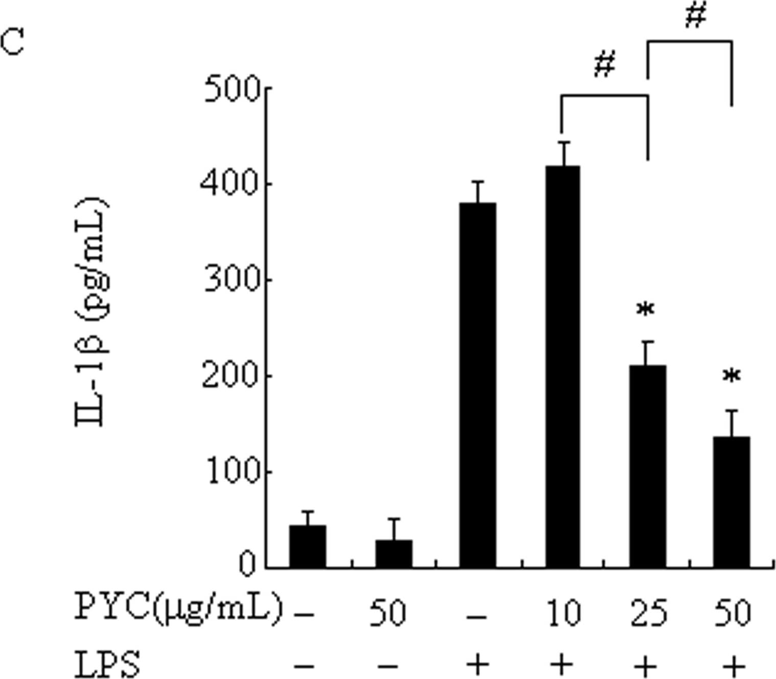
Human E‑Cadherin Antibody PYC suppressed LPS-induced proinflammatory cytokine production in BV2 microglia.Cells were incubated with the indicated concentrations of PYC or vehicle for 1h before LPS treatment (500ng/mL). After 24 h incubation, the culture supernatants were collected, and the amount of TNF-alpha, IL-6, IL-1 beta and IL-10 were measured by ELISA (A-D). Data are represented as mean ± SD from at least 3 independent experiments. *P< 0.05 compared with LPS alone. #P< 0.05. Image collected and cropped by CiteAb from the following open publication (https://dx.plos.org/10.1371/journal.pone.0137837), licensed under a CC-BY license. Not internally tested by R&D Systems.
Product Datasheets
Preparation and Storage
Background: IL-1 beta/IL-1F2
The Interleukin 1 (IL-1) family of proteins consists of IL-1 alpha, IL-1 beta, and the IL-1 receptor antagonist (IL-1ra). IL-1 alpha and IL-1 beta bind to the same cell surface receptors and share biological functions (1). IL-1 is not produced by unstimulated cells of healthy individuals with the exception of skin keratinocytes, some epithelial cells, and certain cells of the central nervous system. However, in response to inflammatory agents, infections, or microbial endotoxins, a dramatic increase in the production of IL-1 by macrophages and various other cell types is seen. IL-1 beta plays a central role in immune and inflammatory responses, bone remodeling, fever, carbohydrate metabolism, and GH/IGF-I physiology. Inappropriate or prolonged production of IL-1 has been implicated in a variety of pathological conditions including sepsis, rheumatoid arthritis, inflammatory bowel disease, acute and chronic myelogenous leukemia, insulindependent diabetes mellitus, atherosclerosis, neuronal injury, and aging-related diseases (2-5).
Background: IL-1 beta/IL-1F2
The Interleukin 1 (IL-1) family of proteins consists of IL-1 alpha, IL-1 beta, and the IL-1 receptor antagonist (IL-1ra). IL-1 alpha and IL-1 beta bind to the same cell surface receptors and share biological functions (1). IL-1 is not produced by unstimulated cells of healthy individuals with the exception of skin keratinocytes, some epithelial cells, and certain cells of the central nervous system. However, in response to inflammatory agents, infections, or microbial endotoxins, a dramatic increase in the production of IL-1 by macrophages and various other cell types is seen. IL-1 beta plays a central role in immune and inflammatory responses, bone remodeling, fever, carbohydrate metabolism, and GH/IGF-I physiology. Inappropriate or prolonged production of IL-1 has been implicated in a variety of pathological conditions including sepsis, rheumatoid arthritis, inflammatory bowel disease, acute and chronic myelogenous leukemia, insulindependent diabetes mellitus, atherosclerosis, neuronal injury, and aging-related diseases (2-5).
