The M-A251 monoclonal antibody specifically binds to the 55 kDa type I transmembrane glycoprotein known as low-affinity interleukin-2 receptor alpha chain subunit (IL-2Rα). CD25 is expressed on regulatory T cells, activated lymphocytes (T and B), and monocytes. It associates with the IL-2Rβ/CD122 and IL-2Rγ/CD132 receptor chains to form the high-affinity IL-2R complex. CD25 expression on T and B lymphocytes is upregulated by antigenic or mitogenic stimulation. Soluble CD25/IL-2Rα is produced as a consequence of lymphocyte stimulation and is found in biological fluids following inflammatory responses.
商品描述
M-A251
The M-A251 monoclonal antibody specifically binds to the 55 kDa type I transmembrane glycoprotein known as low-affinity interleukin-2 receptor alpha chain subunit (IL-2Rα). CD25 is expressed on regulatory T cells, activated lymphocytes (T and B), and monocytes. It associates with the IL-2Rβ/CD122 and IL-2Rγ/CD132 receptor chains to form the high-affinity IL-2R complex. CD25 expression on T and B lymphocytes is upregulated by antigenic or mitogenic stimulation. Soluble CD25/IL-2Rα is produced as a consequence of lymphocyte stimulation and is found in biological fluids following inflammatory responses.
同种型
Mouse BALB/c IgG1, κ
克隆号
克隆 M-A251 (RUO)
产品详情
RB744
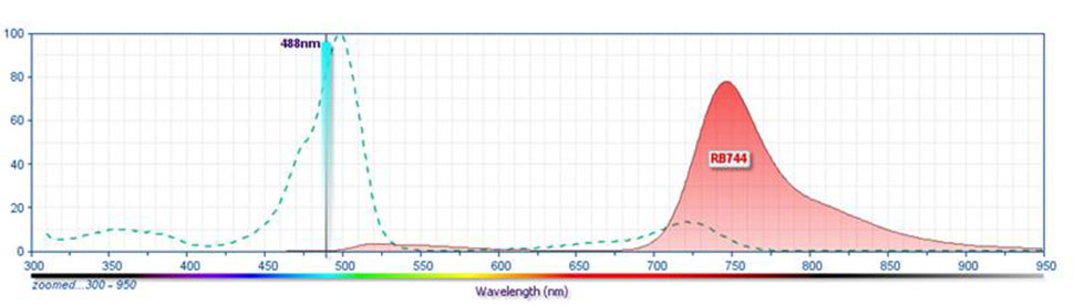
The BD Horizon RealBlue™ 744 (RB744) Dye is part of the BD® family of blue dyes. It is a tandem fluorochrome with an excitation maximum (Ex Max) at 498-nm and an emission maximum (Em Max) at 746-nm as measured using an antibody-dye conjugate. Driven by BD® innovation, RB744 can be used on both spectral and conventional cytometers and is designed to be excited by the Blue laser (488-nm) with minimal excitation by the 561-nm Yellow-Green laser. For conventional instruments equipped with a Blue laser (488-nm), we recommend using an optical filter centered near 750-nm (e.g., a 750/60-nm bandpass filter).
RB744
Blue 488 nm
498 nm
746 nm
应用
实验应用
Flow cytometry (Routinely Tested)
推荐用量
5 µl/test
反应种属
Human (QC Testing), Rhesus,Cynomolgus,Baboon (Tested in Development)
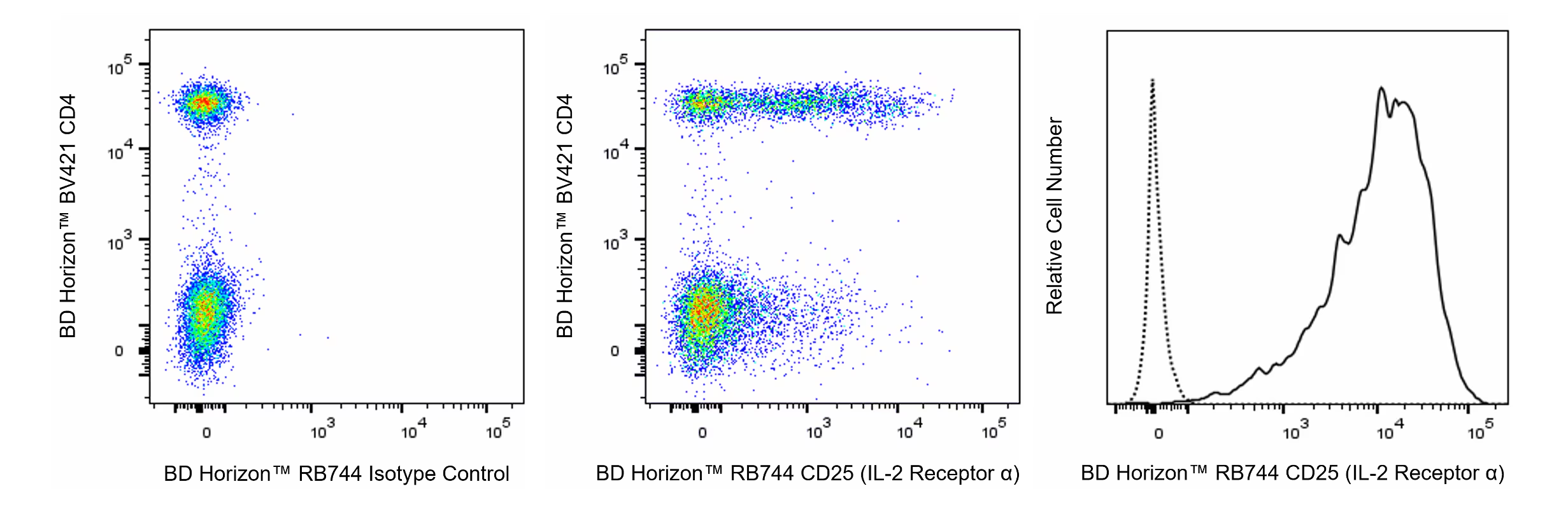
Flow cytometric analysis of CD25 (IL-2 Receptor α) expression on unstimulated and PHA-stimulated Human peripheral blood lymphocytes Left and Middle Plots - Human whole blood was treated with BD Pharm Lyse™ Lysing Buffer (Cat. No. 555899) to lyse erythrocytes. The leukocytes were washed, preincubated with BD Pharmingen™ Human BD Fc Block™ (Cat. No. 564219) and stained with BD Horizon™ BV421 Mouse Anti-Human CD4 antibody (Cat. No. 562424/562425) and with either BD Horizon™ RB744 Mouse IgG1, κ Isotype Control (Cat. No. 570519; Left Plot) or BD Horizon™ RB744 Mouse Anti-Human CD25 (IL-2 Receptor α) antibody (Cat. No. 570620/570708; Middle Plot). The bivariate pseudocolor density plot showing the correlated expression of CD25 (IL-2 Receptor α) [or Ig Isotype control staining] versus CD4 was derived from gated events with the forward and side light-scatter characteristics of viable lymphocytes. Right Plot - Human peripheral blood mononuclear cells were stimulated for 3 days with Phytohemagglutinin (PHA). The cells were stained with either BD Horizon™ RB744 Mouse IgG1, κ Isotype Control (dashed line histogram) or BD Horizon™ RB744 Mouse Anti-Human CD25 (IL-2 Receptor α) antibody (solid line histogram). DAPI (4',6-Diamidino-2-Phenylindole, Dihydrochloride) Solution (Cat. No. 564907) was added to cells right before analysis. The fluorescence histogram showing CD25 (IL-2 Receptor α) expression (or Ig Isotype control staining) was derived from gated events with the forward and side light-scatter characteristics of viable (DAPI-negative) lymphoblasts. Flow cytometry and data analysis were performed using a BD FACSymphony™ A5 SE Cell Analyzer System and FlowJo™ Software.


 全部商品分类


 下载产品说明书
 用小程序,查商品更便捷


 收藏
收藏
 对比
对比 咨询
咨询