The 7G3 monoclonal antibody specifically recognizes human CD123, the 70 kDa IL-3 Receptor α (IL-3Rα) chain. CD123 associates with CD131, the 120-140 kDa Common β chain to form the IL-3 Receptor Complex. CD131 is shared with the receptors for interleukins IL-5 and GM-CSF. IL-3Rα is expressed on hematopoietic progenitors and plays an important role in hematopoietic progenitor cell growth and differentiation. It is also expressed by mast cells, macrophages and a CD5+ B cell subset. This antibody has been reported to block the binding of 125I-IL-3 to high and low affinity IL-3 receptors. In functional experiments, this antibody was found to inhibit acute myeloid leukemia cell proliferation, basophil histamine release, endothelial cell-mediated IL-8 secretion, and neutrophil transmigration. This antibody has been reported to be useful for immunoprecipitation, Western blot and immunofluorescent staining for flow cytometry. At the Fifth HLDA Workshop, the human IL-3 receptor was designated CD123.
商品描述
7G3
The 7G3 monoclonal antibody specifically recognizes human CD123, the 70 kDa IL-3 Receptor α (IL-3Rα) chain. CD123 associates with CD131, the 120-140 kDa Common β chain to form the IL-3 Receptor Complex. CD131 is shared with the receptors for interleukins IL-5 and GM-CSF. IL-3Rα is expressed on hematopoietic progenitors and plays an important role in hematopoietic progenitor cell growth and differentiation. It is also expressed by mast cells, macrophages and a CD5+ B cell subset. This antibody has been reported to block the binding of 125I-IL-3 to high and low affinity IL-3 receptors. In functional experiments, this antibody was found to inhibit acute myeloid leukemia cell proliferation, basophil histamine release, endothelial cell-mediated IL-8 secretion, and neutrophil transmigration. This antibody has been reported to be useful for immunoprecipitation, Western blot and immunofluorescent staining for flow cytometry. At the Fifth HLDA Workshop, the human IL-3 receptor was designated CD123.
同种型
Mouse IgG2a, κ
克隆号
克隆 7G3 (RUO)
浓度
0.2 mg/ml
产品详情
PE
R-Phycoerythrin (PE), is part of the BD family of Phycobiliprotein dyes. This fluorochrome is a multimeric fluorescent phycobiliprotein with excitation maximum (Ex Max) of 496 nm and 566 nm and an emission maximum (Em Max) at 576 nm. PE is designed to be excited by the Blue (488 nm), Green (532 nm) and Yellow-Green (561 nm) lasers and detected using an optical filter centered near 575 nm (e.g., a 575/26-nm bandpass filter). As PE is excited by multiple lasers, this can result in cross-laser excitation and fluorescence spillover on instruments with various combinations of Blue, Green, and Yellow-Green lasers. Please ensure that your instrument’s configurations (lasers and optical filters) are appropriate for this dye.
Aqueous buffered solution containing protein stabilizer and ≤0.09% sodium azide.
保存方式
Aqueous buffered solution containing protein stabilizer and ≤0.09% sodium azide.
数据库链接
Entrez-Gene ID
3563
参考图片
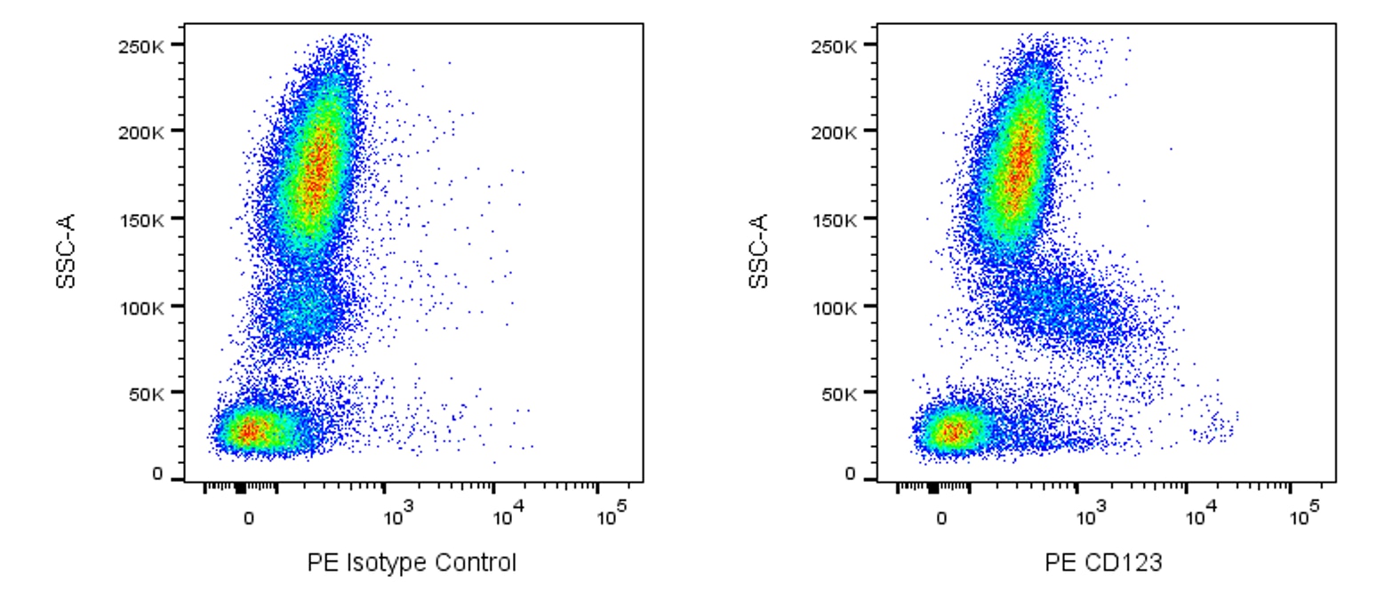
Peripheral blood lymphocytes analyzed on a FACScan (BDIS, San Jose, CA)
Multiparameter flow cytometric analysis of CD123 expression on human peripheral blood leucocyte populations. Human whole blood was stained with either PE Mouse IgG2a, κ Isotype Control (Cat. No. 554648; Left Plot) or PE Mouse Anti-Human CD123 antibody (Cat. No. 554529/561058; Right Plot) at 0.5 µg/test. Erythrocytes were lysed with BD Pharm Lyse™ Lysing Solution (Cat. No. 555899). Bivariate pseudocolor density plots showing the correlated expression of CD123 (IL-3 Receptor α) [or Ig Isotype control staining] versus side light-scatter (SSC-A) signals were derived from gated events with the forward and side light-scatter characteristics of intact leucocyte populations. Flow cytometry and data analysis was performed using a BD LSRFortessa™ Cell Analyzer System and FlowJo™ software. Data shown on this Technical Data Sheet are not lot specific.
Peripheral blood lymphocytes analyzed on a FACScan (BDIS, San Jose, CA)


 全部商品分类


 用小程序,查商品更便捷


