


 全部商品分类
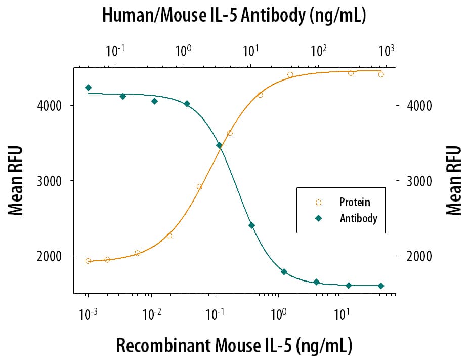


 下载产品说明书 下载SDS
 用小程序,查商品更便捷


 收藏
收藏
 对比
对比 咨询
咨询
Scientific Data
 View Larger
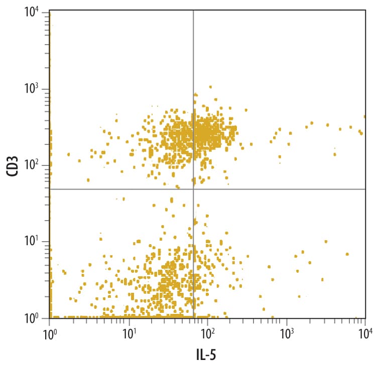
Detection of IL‑5 in Human PBMCs by Flow Cytometry. Human peripheral blood monocytes treated with 50 ng/mL PMA and 500 ng/mL Calcium Ionomycin for 6 hours were stained with Rat Anti-Human/Mouse IL-5 Monoclonal Antibody (Catalog # MAB405) followed by Phycoerythrin-conjugated Anti-Rat IgG F(ab')2Secondary Antibody (Catalog # F0105B) and Human CD3e APC-conjugated Monoclonal Antibody (Catalog # FAB100A). Quadrant markers were set based on control antibody staining (Catalog # MAB005). To facilitate intracellular staining, cells were fixed with paraformaldehyde and permeabilized with saponin.
 View Larger
Cell Proliferation Induced by IL‑5 and Neutralization by Human/Mouse IL‑5 Antibody. Recombinant Mouse IL-5 (Catalog # 405-ML) stimulates proliferation in the TF-1 human erythroleukemic cell line in a dose-dependent manner (orange line). Proliferation elicited by Recombinant Mouse IL-5 (0.5 ng/mL) is neutralized (green line) by increasing concentrations of Rat Anti-Human/Mouse IL-5 Monoclonal Antibody (Catalog # MAB405). The ND50 is typically 0.004-0.015 µg/mL.
Human/Mouse IL-5 Antibody Summary
Applications
Mouse IL-5 Sandwich Immunoassay
Human IL-5 Sandwich Immunoassay
Please Note: Optimal dilutions should be determined by each laboratory for each application. General Protocols are available in the Technical Information section on our website.
Background: IL-5
IL-5 is a disulfide-linked homodimeric cytokine that is secreted by T cells. IL-5 promotes the proliferation, differentation, and activation of eosinophils. It binds to a receptor complex consisting of one IL-5 specific alpha chain and one non-binding common beta chain that is shared with the receptors for GM-CSF and IL-3.
Preparation and Storage
- 12 months from date of receipt, -20 to -70 °C as supplied.
- 1 month, 2 to 8 °C under sterile conditions after reconstitution.
- 6 months, -20 to -70 °C under sterile conditions after reconstitution.
参考图片
Detection of IL‑5 in Human PBMCs by Flow Cytometry. Human peripheral blood monocytes treated with 50 ng/mL PMA and 500 ng/mL Calcium Ionomycin for 6 hours were stained with Rat Anti-Human/Mouse IL‑5 Monoclonal Antibody (Catalog # MAB405) followed by Phycoerythrin-conjugated Anti-Rat IgG F(ab')2 Secondary Antibody (Catalog # F0105B) and Human CD3 epsilon APC-conjugated Monoclonal Antibody (Catalog # FAB100A). Quadrant markers were set based on control antibody staining (Catalog # MAB005). To facilitate intracellular staining, cells were fixed with paraformaldehyde and permeabilized with saponin.
Cell Proliferation Induced by IL‑5 and Neutralization by Human/Mouse IL‑5 Antibody. Recombinant Mouse IL‑5 (Catalog # 405-ML) stimulates proliferation in the TF‑1 human erythroleukemic cell line in a dose-dependent manner (orange line). Proliferation elicited by Recombinant Mouse IL‑5 (0.5 ng/mL) is neutralized (green line) by increasing concentrations of Rat Anti-Human/Mouse IL‑5 Monoclonal Antibody (Catalog # MAB405). The ND50 is typically
0.004-0.015 µg/mL.