


 全部商品分类
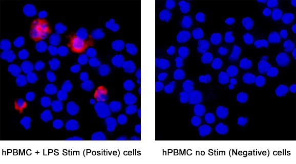


 下载产品说明书 下载SDS
 用小程序,查商品更便捷


 收藏
收藏
 对比
对比 咨询
咨询
Val30-Met212
Accession # P05231
Scientific Data
.") View Larger
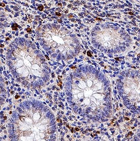
View LargerIL‑6 in Human Appendix. IL-6 was detected in immersion fixed paraffin-embedded sections of human appendix using Mouse Anti-Human IL-6 Monoclonal Antibody (Catalog # MAB2061) at 1.7 µg/mL for 1 hour at room temperature followed by incubation with the Anti-Mouse IgG VisUCyte™ HRP Polymer Antibody (VC001). Before incubation with the primary antibody, tissue was subjected to heat-induced epitope retrieval using Antigen Retrieval Reagent-Basic (CTS013). Tissue was stained using DAB (brown) and counterstained with hematoxylin (blue). Specific staining was localized to cytoplasm in lymphocytes. View our protocol for IHC Staining with VisUCyte HRP Polymer Detection Reagents.
 View Larger
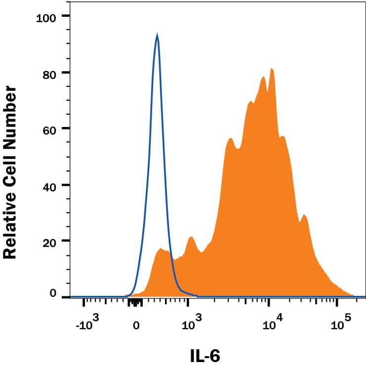
Detection of IL‑6 in Human PBMCs by Flow Cytometry. Human peripheral blood mononuclear cells (PBMCs) treated with 100 ng/mL LPS for 24 hours were stained with Mouse Anti-Human IL-6 Monoclonal Antibody (Catalog # MAB2061, filled histogram) or isotype control antibody (MAB004, open histogram), followed by Phycoerythrin-conjugated Anti-Mouse IgG Secondary Antibody (F0102B). To facilitate intracellular staining, cells were fixed with Flow Cytometry Fixation Buffer (FC004) and permeabilized with Flow Cytometry Permeabilization/Wash Buffer I (FC005). View our protocol for Staining Intracellular Molecules.
 View Larger
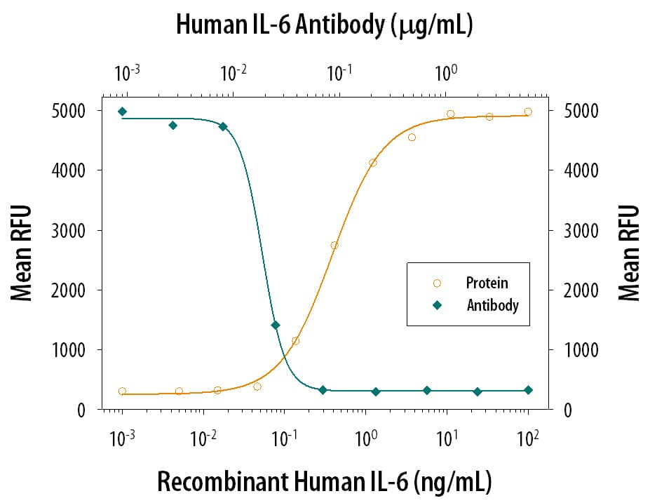
Cell Proliferation Induced by IL‑6 and Neutralization by Human IL‑6 Antibody. Recombinant Human IL-6 (206-IL) stimulates proliferation in the T1165.85.2.1 mouse plasmacytoma cell line in a dose-dependent manner (orange line). Proliferation elicited by Recombinant Human IL-6 (2.5 ng/mL) is neutralized (green line) by increasing concentrations of Mouse Anti-Human IL-6 Monoclonal Antibody (Catalog # MAB2061). The ND50 is typically 8.00-80.0 ng/mL.
 View Larger
Detection of IL‑6 in Human PBMCs. IL‑6 was detected in immersion fixed Human PBMCs stimulated with LPS (positive control) and absent in untreated human PBMCs (negative control) using Mouse Anti-Human IL‑6 Monoclonal Antibody (Catalog # MAB2061) at 3 µg/mL for 3 hours at room temperature. Cells were stained using the NorthernLights™ 557-conjugated Anti-Mouse IgG Secondary Antibody (red; Catalog # NL007) and counterstained with DAPI (blue). Specific staining was localized to cytoplasm. View our protocol for Fluorescent ICC Staining of Non-adherent Cells.
 View Larger
Detection of IL‑6 in PBMC monocytes by Flow Cytometry. PBMCs stimulated with 100 ng/ml LPS and Brefeldin A for 24 hrs (A) or unstimulated (B) were stained with Mouse Anti-Human IL‑6 Monoclonal Antibody (Catalog # MAB2061) and Mouse Anti-Human CD14 PE‑conjugated Monoclonal Antibody (Catalog # FAB3832P). To facilitate intracellular staining, cells were fixed with Flow Cytometry Fixation Buffer (1X) (Catalog # FC004) and permeabilized with Saponin. View our protocol for Staining Intracellular Molecules.
Human IL-6 Antibody Summary
Val30-Met212
Accession # P05231
*Small pack size (-SP) is supplied either lyophilized or as a 0.2 µm filtered solution in PBS.
Applications
Please Note: Optimal dilutions should be determined by each laboratory for each application. General Protocols are available in the Technical Information section on our website.
Background: IL-6
Interleukin-6 (IL-6) is a pleiotropic, alpha -helical, phosphorylated and variably glycosylated cytokine that plays important roles in the acute phase reaction, inflammation, hematopoiesis, bone metabolism, and cancer progression. Mature human IL-6 is 183 amino acids (aa) in length expressed as a 22-28 kDA molecular weight protein. IL-6 shares 39% aa sequence identity with mouse and rat IL-6. Alternative splicing generates several isoforms with internal deletions, some of which exhibit antagonistic properties. IL-6 induces signaling through a cell surface heterodimeric receptor complex composed of a ligand binding subunit (IL-6 R alpha) and a signal transducing subunit (gp130). IL-6 binds to IL-6 R alpha, triggering IL-6 R alpha association with gp130 and gp130 dimerization. gp130 is also a component of the receptors for CLC, CNTF, CT-1, IL-11, IL-27, LIF, and OSM. Soluble forms of IL-6 R alpha are generated by both alternative splicing and proteolytic cleavage. In a mechanism known as trans-signaling, complexes of soluble IL-6 and IL-6 R alpha elicit responses from gp130-expressing cells that lack cell surface IL-6 R alpha. Trans-signaling enables a wider range of cell types to respond to IL-6, as the expression of gp130 is ubiquitous, while that of IL-6 R alpha is predominantly restricted to hepatocytes, monocytes, and resting lymphocytes. Soluble splice forms of gp130 block trans-signaling from IL-6/IL-6 R alpha but not from other cytokines that use gp130 as a co-receptor. IL-6, along with TNF-alpha and IL-1, function to drive the acute inflammatory response and the transition from acute inflammation to either acquired immunity or chronic inflammatory disease. When dysregulated, it contributes to chronic inflammation in obesity, insulin resistance, inflammatory bowel disease, arthritis, sepsis, and atherosclerosis. IL-6 can also function as an anti-inflammatory molecule, as in skeletal muscle where it is secreted in response to exercise. In addition, it enhances hematopoietic stem cell proliferation and the differentiation of Th17 cells, memory B cells, and plasma cells.
Preparation and Storage
- 12 months from date of receipt, -20 to -70 °C as supplied.
- 1 month, 2 to 8 °C under sterile conditions after reconstitution.
- 6 months, -20 to -70 °C under sterile conditions after reconstitution.
参考图片
IL‑6 in Human PBMCs. IL‑6 was detected in immersion fixed human peripheral blood mononuclear cells (PBMCs) stimulated with LPS and monensin using Mouse Anti-Human IL‑6 Monoclonal Antibody (Catalog # MAB2061) at 10 µg/mL for 3 hours at room temperature. Cells were stained using the NorthernLights™ 557-conjugated Anti-Mouse IgG Secondary Antibody (yellow; Catalog # NL007) and counterstained with DAPI (blue). View our protocol for Fluorescent ICC Staining of Non-adherent Cells.
Cell Proliferation Induced by IL‑6 and Neutralization by Human IL‑6 Antibody. Recombinant Human IL‑6 (Catalog # 206-IL) stimulates proliferation in the T1165.85.2.1 mouse plasmacytoma cell line in a dose-dependent manner (orange line). Proliferation elicited by Recombinant Human IL‑6 (2.5 ng/mL) is neutralized (green line) by increasing concentrations of Mouse Anti-Human IL‑6 Monoclonal Antibody (Catalog # MAB2061). The ND50 is typically 0.05-0.15 µg/mL.
Detection of IL‑6 in Human PBMCs by Flow Cytometry. Human peripheral blood mononuclear cells (PBMCs) treated with
100 μg/mL LPS for 24 hours were stained with Mouse Anti-Human IL‑6 Monoclonal Antibody (Catalog # MAB2061, filled histogram) or isotype control antibody (Catalog # MAB004, open histogram), followed by Phycoerythrin-conjugated Anti-Mouse IgG Secondary Antibody (Catalog # F0102B). To facilitate intracellular staining, cells were fixed with Flow Cytometry Fixation Buffer (Catalog # FC004) and permeabilized with Flow Cytometry Permeabilization/Wash Buffer I (Catalog # FC005). View our protocol for Staining Intracellular Molecules.