


 全部商品分类
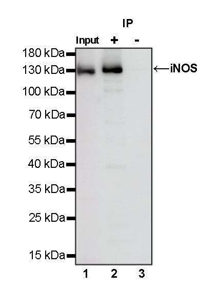


iNOS Recombinant Rabbit mAb (S-466-102)

 下载产品说明书
 用小程序,查商品更便捷


 收藏
收藏
 对比
对比 咨询
咨询
WB
1:1000IHC-P
1:100ICC
1:500IP
1:50ICFCM
1:500
Nitric oxide synthases (NOSs) are a family of enzymes catalyzing the production of nitric oxide (NO) from L-arginine. NO is an important cellular signaling molecule. It helps modulate vascular tone, insulin secretion, airway tone, and peristalsis, and is involved in angiogenesis and neural development. It may function as a retrograde neurotransmitter. Nitric oxide is mediated in mammals by the calcium-calmodulin controlled isoenzymes eNOS (endothelial NOS) and nNOS (neuronal NOS). The inducible isoform, iNOS, involved in immune response, binds calmodulin at physiologically relevant concentrations, and produces NO as an immune defense mechanism.
12 months from date of receipt / reconstitution, -20 °C as supplied
参考图片
WB result of iNOS Rabbit mAb
Primary antibody: iNOS Rabbit mAb at 1/1000 dilution
Lane 1: RAW 264.7 whole cell lysate 20 µg
Lane 2: RAW 264.7 treated with LPS (1 µg/mL, 16 hr) whole cell lysate 20 µg
Secondary antibody: Goat Anti-Rabbit IgG, (H+L), HRP conjugated at 1/10000 dilution
Predicted MW: 130 kDa
Observed MW: 130 kDa
IHC shows positive staining in paraffin-embedded mouse lung. Anti-iNOS antibody was used at 1/100 dilution, followed by a HRP Polymer for Mouse & Rabbit IgG (ready to use). Counterstained with hematoxylin. Heat mediated antigen retrieval with Tris/EDTA buffer pH9.0 was performed before commencing with IHC staining protocol.
IHC shows positive staining in paraffin-embedded rat lung. Anti-iNOS antibody was used at 1/100 dilution, followed by a HRP Polymer for Mouse & Rabbit IgG (ready to use). Counterstained with hematoxylin. Heat mediated antigen retrieval with Tris/EDTA buffer pH9.0 was performed before commencing with IHC staining protocol.
ICC shows positive staining in Raw264.7 cells. Anti-iNOS antibody was used at 1/500 dilution (Green) and incubated overnight at 4°C. Goat polyclonal Antibody to Rabbit IgG - H&L (Alexa Fluor® 488) was used as secondary antibody at 1/1000 dilution. The cells were fixed with 100% ice-cold methanol and permeabilized with 0.1% PBS-Triton X-100. Nuclei were counterstained with DAPI (Blue). Counterstain with tubulin (Red).
iNOS Rabbit mAb at 1/50 dilution (1 µg) immunoprecipitating iNOS in 0.4 mg RAW 264.7 treated with LPS (1 µg/mL, 16 hr) whole cell lysate.
Western blot was performed on the immunoprecipitate using iNOS Rabbit mAb at 1/1000 dilution.
Secondary antibody (HRP) for IP was used at 1/400 dilution.
Lane 1: RAW 264.7 treated with LPS (1 µg/mL, 16 hr) whole cell lysate 20 µg (Input)
Lane 2: iNOS Rabbit mAb IP in RAW 264.7 treated with LPS (1 µg/mL, 16 hr) whole cell lysate
Lane 3: Rabbit monoclonal IgG IP in RAW 264.7 treated with LPS (1 µg/mL, 16 hr) whole cell lysate
Predicted MW: 130 kDa
Observed MW: 130 kDa
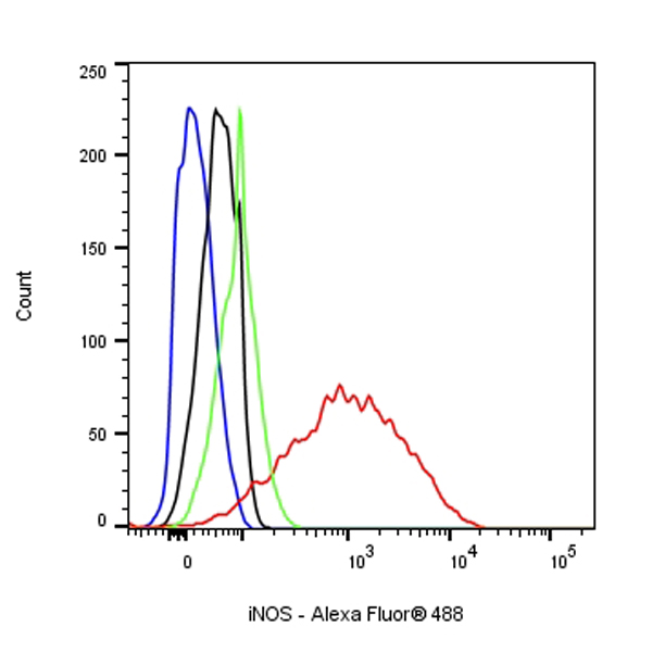
Flow cytometric analysis of 4% PFA fixed 90% methanol permeabilized RAW264.7 (Mouse Abelson murine leukemia virus-induced tumor macrophage), treated with 1μg/ml LPS for 16h (Red) or untreated (Green), labeling iNOS at 1/500 dilution (0.1 μg) compared with a Rabbit monoclonal IgG isotype control (Black) and an unlabeled control (cells without incubation with primary antibody and secondary antibody) (Blue). Goat Anti - Rabbit IgG Alexa Fluor® 488 was used as the secondary antibody.