


 用小程序,查商品更便捷


WB
1:1000IP
1:25IHC-P
1:500ICC
1:500ICFCM
1:500
CD11b, also known as Integrated alpha-m, a transgender protein, can form an heterododerous composed of α and β subunit. It is a common bone marrow mark (neutral granulocyte, monocyte, macrophage, and small gel cells) and NK (natural kill cells) antigens. It can be used to distinguish between acute granulocyte deficiency (CD11B+, CD117--) and acute early early early elastic cell leukemia (CD11B-, CD117+).
12 months from date of receipt / reconstitution, -20 °C as supplied
参考图片
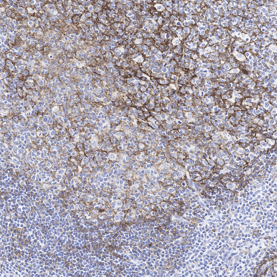
IHC shows positive staining in paraffin-embedded human tonsil
.Anti-CD11b antibody was used at 1/500 dilution, followed by a Goat Anti-Rabbit IgG H&L (HRP) ready to use. Counterstained with hematoxylin.
Heat mediated antigen retrieval with Tris/EDTA buffer pH9.0 was performed before commencing with IHC staining protocol.
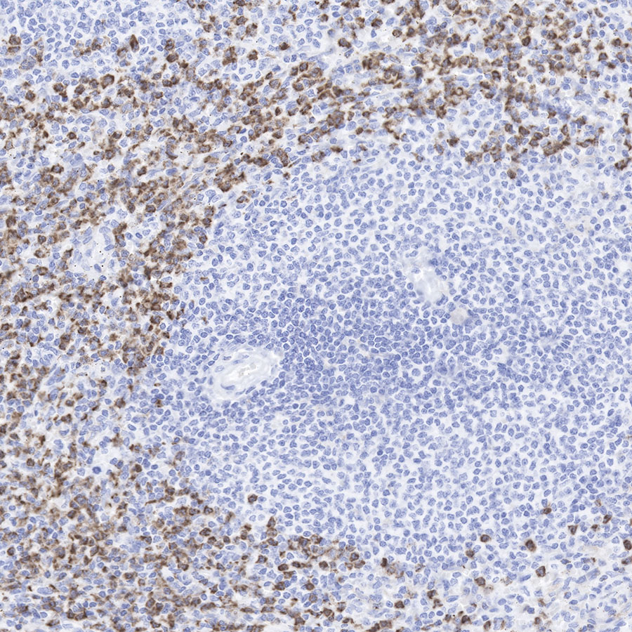
IHC shows positive staining in paraffin-embedded human spleen.
Anti-CD11b antibody was used at 1/500 dilution, followed by a Goat Anti-Rabbit IgG H&L (HRP) ready to use. Counterstained with hematoxylin.
Heat mediated antigen retrieval with Tris/EDTA buffer pH9.0 was performed before commencing with IHC staining protocol.
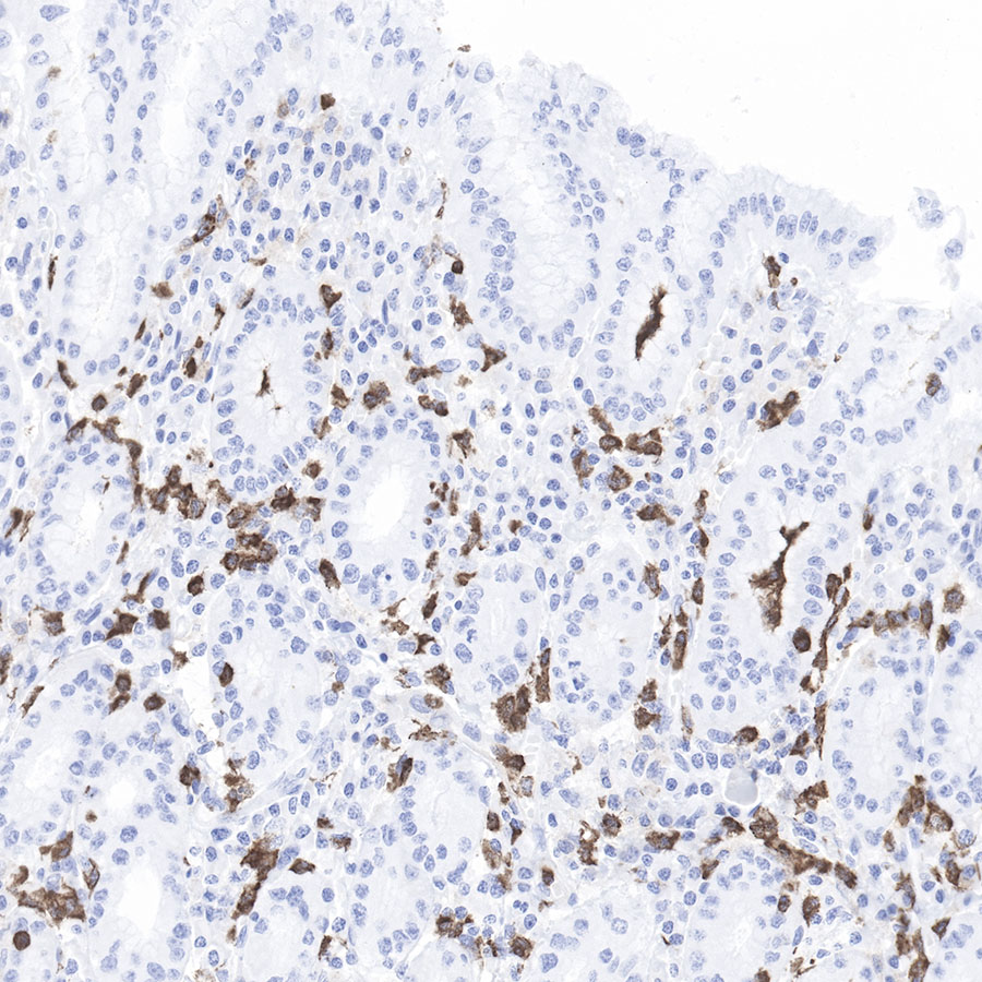
IHC shows positive staining in paraffin-embedded human stomach.
Anti-CD11b antibody was used at 1/500 dilution, followed by a Goat Anti-Rabbit IgG H&L (HRP) ready to use. Counterstained with hematoxylin.
Heat mediated antigen retrieval with Tris/EDTA buffer pH9.0 was performed before commencing with IHC staining protocol.
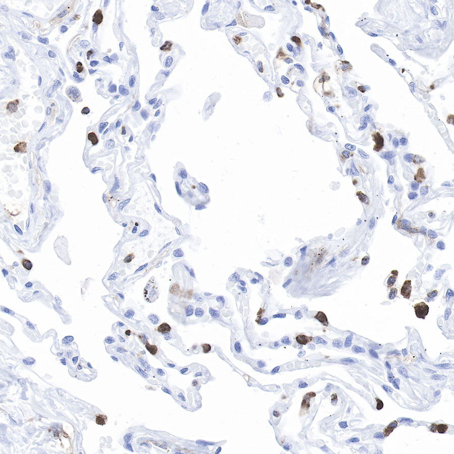
IHC shows positive staining in paraffin-embedded human lung.
Anti-CD11b antibody was used at 1/500 dilution, followed by a Goat Anti-Rabbit IgG H&L (HRP) ready to use. Counterstained with hematoxylin.
Heat mediated antigen retrieval with Tris/EDTA buffer pH9.0 was performed before commencing with IHC staining protocol.
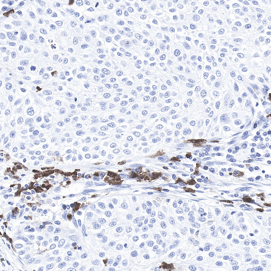
IHC shows positive staining in paraffin-embedded human cervix cancer.
Anti-CD11b antibody was used at 1/500 dilution, followed by a Goat Anti-Rabbit IgG H&L (HRP) ready to use. Counterstained with hematoxylin.
Heat mediated antigen retrieval with Tris/EDTA buffer pH9.0 was performed before commencing with IHC staining protocol.
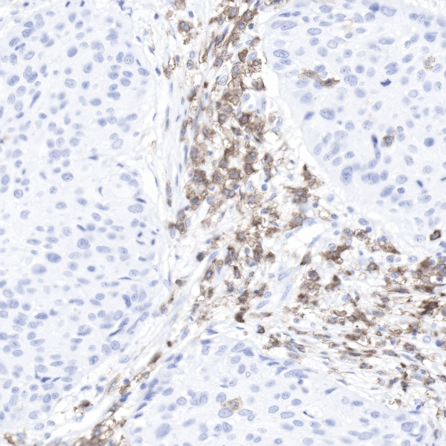
IHC shows positive staining in paraffin-embedded human lung squamous cancer.
Anti-CD11b antibody was used at 1/500 dilution, followed by a Goat Anti-Rabbit IgG H&L (HRP) ready to use. Counterstained with hematoxylin.
Heat mediated antigen retrieval with Tris/EDTA buffer pH9.0 was performed before commencing with IHC staining protocol.
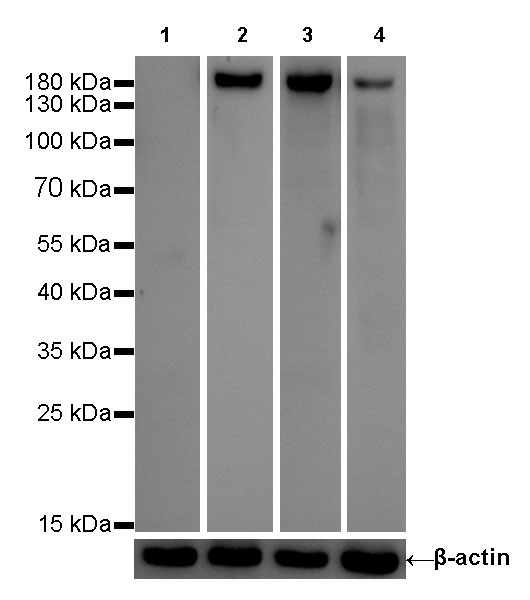
WB result of CD11b Rabbit mAb
Primary antibody:CD11b Rabbit mAb at 1/1000 dilution
Lane 1: Jurkat whole cell lysate 20 µg
Lane 2: U937 whole cell lysate 20 µg
Lane 3: TF-1 whole cell lysate 20 µg
Lane 4: THP-1 whole cell lysate 20 µg
Negative control: Jurkat whole cell lysate
Secondary antibody: Goat Anti-Rabbit IgG, (H+L), HRP conjugated at 1/10000 dilution
Predicted MW: 170 kDa
Observed MW: 180 kDa
Exposure time: Lane 1、lane 2 and lane 4: 180s
Lane 3: 20s
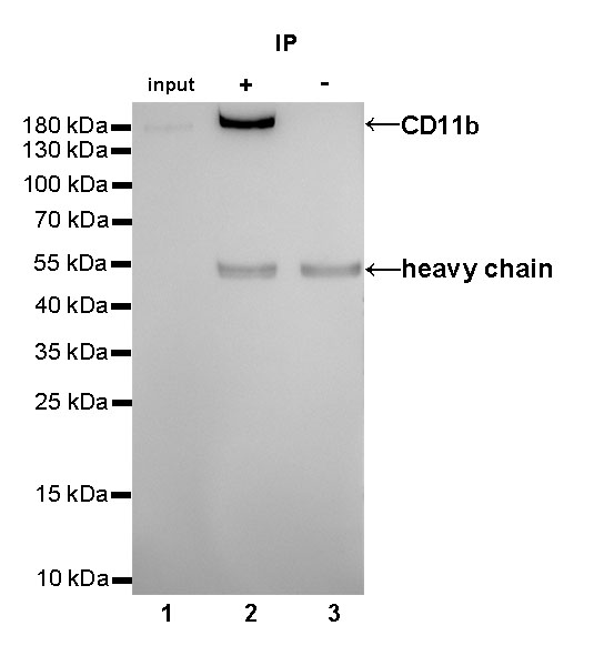
CD11b Rabbit mAb at 1/25 dilution (2µg) immunoprecipitating CD11b in 0.4mg TF-1 whole cell lysate.
Western blot was performed on the immunoprecipitate using CD11b Rabbit mAb at 1/1000 dilution.
Secondary antibody (HRP) for IP was used at 1/400 dilution.
Lane 1: TF-1 whole cell lysate 10µg (input)
Lane 2 (+): CD11b Rabbit mAb IP in TF-1 whole cell lysate
Lane 3 (-): Rabbit monoclonal IgG IP in TF-1 whole cell lysate
Predicted MW: 170 kDa
Observed MW: 180 kDa
Exposure time: 10s
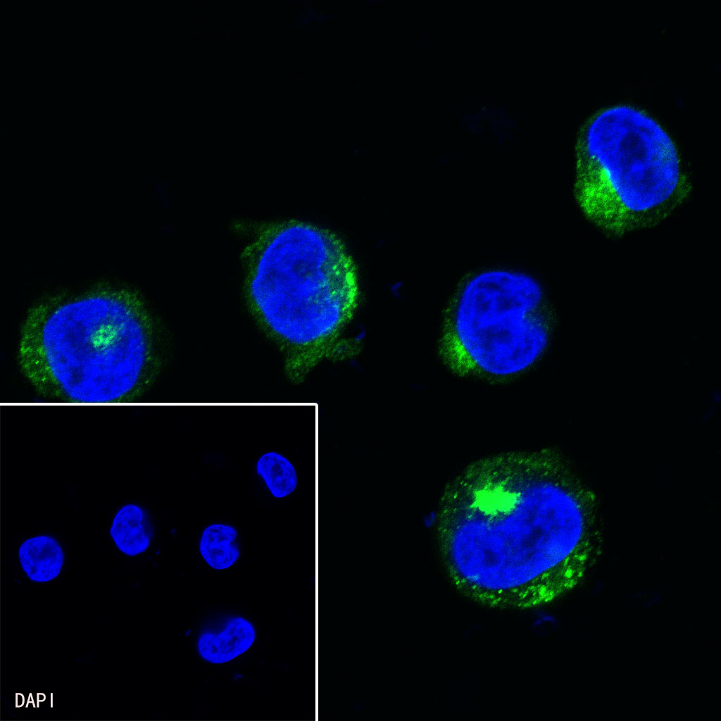
ICC shows positive staining in TF-1 cells. Anti-CD11b antibody was used at 1/500 dilution (Green) and incubated overnight at 4°C. Goat polyclonal Antibody to Rabbit IgG - H&L (Alexa Fluor® 488) was used as secondary antibody at 1/1000 dilution. The cells were fixed with 4%PFA and permeabilized with 0.1% PBS-Triton X-100. Nuclei were counterstained with DAPI (Blue).
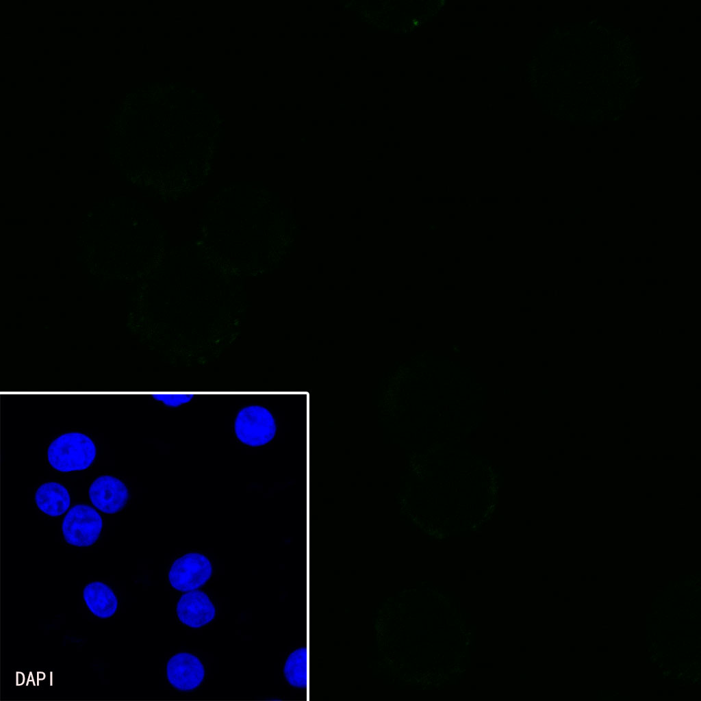
Negative control:ICC shows negative staining in Jurkat cells. Anti-CD11b antibody was used at 1/500 dilution and incubated overnight at 4°C. Goat polyclonal Antibody to Rabbit IgG - H&L (Alexa Fluor® 488) was used as secondary antibody at 1/1000 dilution. The cells were fixed with 4%PFA and permeabilized with 0.1% PBS-Triton X-100. Nuclei were counterstained with DAPI (Blue).
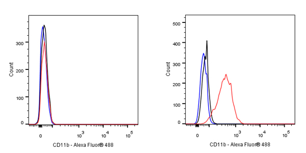
Flow cytometric analysis of 4% PFA fixed 90% methanol permeabilized Jurkat (Human T cell leukemia T lymphocyte, left) / TF-1 (Human Erythroleukemia erythroblast, Right) cells labelling CD11b antibody at 1/500 dilution (0.1 μg) / (Red) compared with a Rabbit monoclonal IgG (Black) isotype control and an unlabelled control (cells without incubation with primary antibody and secondary antibody) (Blue). Goat Anti - Rabbit IgG Alexa Fluor® 488 was used as the secondary antibody.
Negative control: Jurkat

 危险品化学品经营许可证(不带存储) 许可证编号:沪(杨)应急管危经许[2022]202944(QY)
  营业执照(三证合一)