


 全部商品分类


BD Pharmingen™ FITC Hamster Anti-Mouse CD11c

 下载产品说明书 下载SDS
 用小程序,查商品更便捷


 收藏
收藏
 对比
对比 咨询
咨询

参考图片
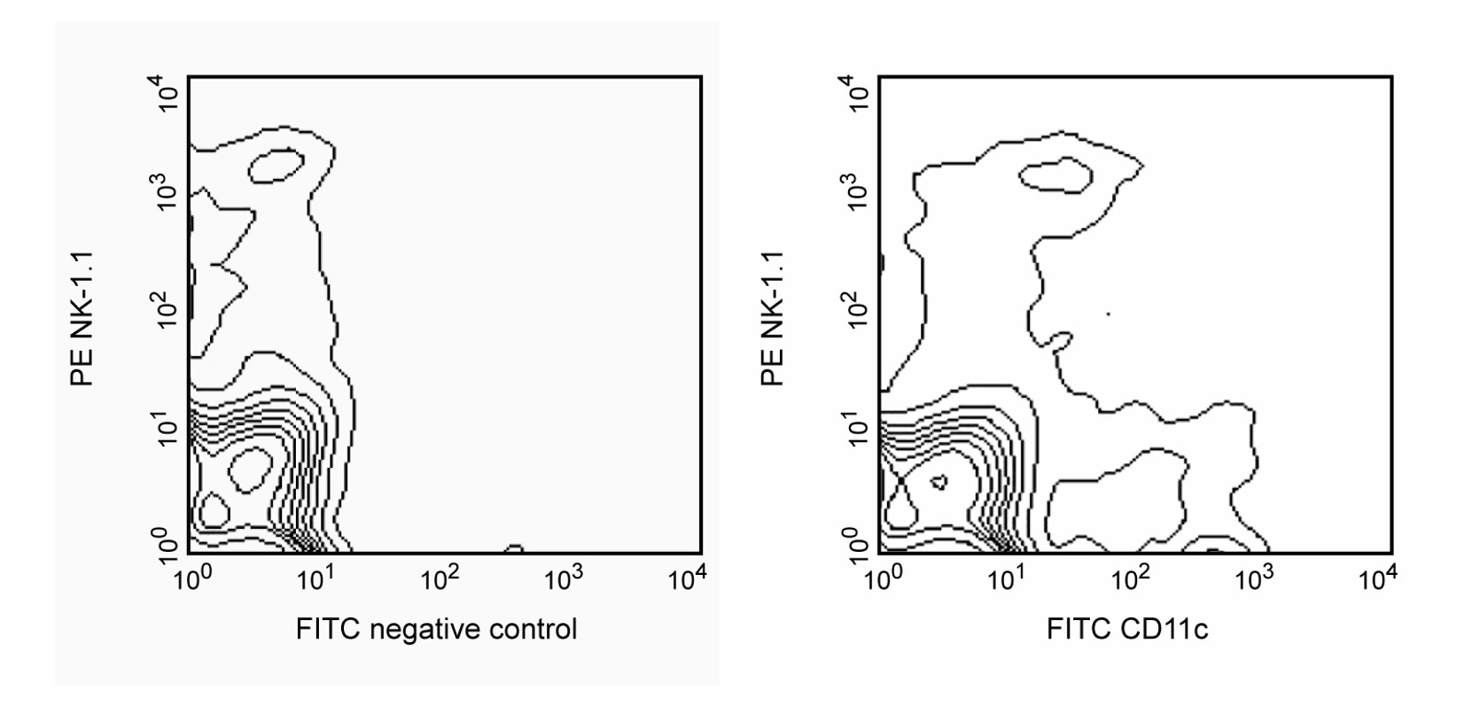
Flow cytometric analysis of CD11c expression on mouse splenocytes. Spleen NK cells were stained with PE Mouse Anti-Mouse NK-1.1 (Cat. No. 557391/553165) and either FITC Hamster IgG Isotype Control (Cat. No. 553953; left panel) or FITC Hamster anti-Mouse CD11c antibody (Cat. No. 553801/557400; right panel). The contour plots were derived from events with the forward and side light-scatter characteristics of viable splenocytes. Flow cytometric analysis was performed using a BD FACSCan™ flow cytometry system.
Flow cytometric analysis of CD11c expression on mouse splenocytes. Spleen NK cells were stained with PE Mouse Anti-Mouse NK-1.1 (Cat. No. 557391/553165) and either FITC Hamster IgG Isotype Control (Cat. No. 553953; left panel) or FITC Hamster anti-Mouse CD11c antibody (Cat. No. 553801/557400; right panel). The contour plots were derived from events with the forward and side light-scatter characteristics of viable splenocytes. Flow cytometric analysis was performed using a BD FACSCan™ flow cytometry system.