The B-ly6 monoclonal antibody specifically binds to the 150 kDa adhesion glycoprotein CD11c (p150, integrin α chain). CD11c is expressed on dendritic cells, monocytes, macrophages, granulocytes, NK cells and subsets of B and T cells. It associates with CD18 to form the CD11c/CD18 complex that binds fibrinogen and has been reported to be a receptor for iC3b and ICAM-1. Reports indicate that CD11c/CD18 plays a role as an adhesion molecule that mediates cellular binding to ligands expressed on stimulated epithelium and endothelium.
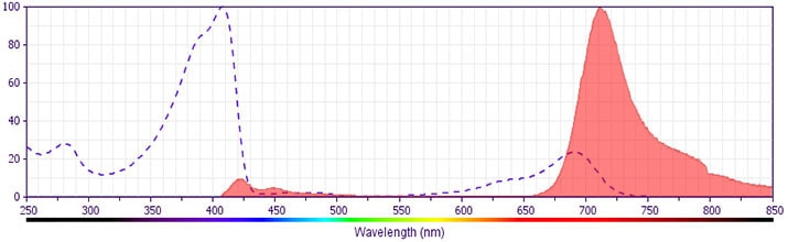
The antibody was conjugated to BD Horizon BV711 which is part of the BD Horizon Brilliant™ Violet family of dyes. This dye is a tandem fluorochrome of BD Horizon BV421 with an Ex Max of 405-nm and an acceptor dye with an Em Max at 711-nm. BD Horizon BV711 can be excited by the violet laser and detected in a filter used to detect Cy™5.5 / Alexa Fluor® 700-like dyes (eg, 712/20-nm filter). Due to the excitation and emission characteristics of the acceptor dye, there may be moderate spillover into the Alexa Fluor® 700 and PerCP-Cy5.5 detectors. However, the spillover can be corrected through compensation as with any other dye combination.
商品描述
B-ly6
The B-ly6 monoclonal antibody specifically binds to the 150 kDa adhesion glycoprotein CD11c (p150, integrin α chain). CD11c is expressed on dendritic cells, monocytes, macrophages, granulocytes, NK cells and subsets of B and T cells. It associates with CD18 to form the CD11c/CD18 complex that binds fibrinogen and has been reported to be a receptor for iC3b and ICAM-1. Reports indicate that CD11c/CD18 plays a role as an adhesion molecule that mediates cellular binding to ligands expressed on stimulated epithelium and endothelium.
The antibody was conjugated to BD Horizon BV711 which is part of the BD Horizon Brilliant™ Violet family of dyes. This dye is a tandem fluorochrome of BD Horizon BV421 with an Ex Max of 405-nm and an acceptor dye with an Em Max at 711-nm. BD Horizon BV711 can be excited by the violet laser and detected in a filter used to detect Cy™5.5 / Alexa Fluor® 700-like dyes (eg, 712/20-nm filter). Due to the excitation and emission characteristics of the acceptor dye, there may be moderate spillover into the Alexa Fluor® 700 and PerCP-Cy5.5 detectors. However, the spillover can be corrected through compensation as with any other dye combination.
同种型
Mouse BALB/c IgG1, κ
克隆号
克隆 B-ly6 (RUO)
产品详情
BV711
The BD Horizon Brilliant Violet™ 711 (BV711) Dye is part of the BD Horizon Brilliant Violet™ family of dyes. This tandem fluorochrome is comprised of a BV421 donor with an excitation maximum (Ex Max) of 407-nm and an acceptor dye with an emission maximum (Em Max) at 713-nm. BV711, driven by BD innovation, is designed to be excited by the violet laser (405-nm) and detected using an optical filter centered near 710-nm (e.g., a 712/20-nm bandpass filter). The acceptor dye can be excited by the Red (628–640-nm) laser resulting in cross-laser excitation and fluorescence spillover. Please ensure that your instrument’s configurations (lasers and optical filters) are appropriate for this dye.
Aqueous buffered solution containing BSA and ≤0.09% sodium azide.
保存方式
Aqueous buffered solution containing BSA and ≤0.09% sodium azide.
文献
文献
研发参考(4)
1. Knapp W. W. Knapp .. et al., ed. Leucocyte typing IV : white cell differentiation antigens. Oxford New York: Oxford University Press; 1989:1-1182.
2. Stacker SA, Springer TA. Leukocyte integrin P150,95 (CD11c/CD18) functions as an adhesion molecule binding to a counter-receptor on stimulated endothelium. J Immunol. 1991; 146(2):648-655. (Clone-specific: ELISA).
3. Visser L, Shaw A, Slupsky J, Vos H, Poppema S. Monoclonal antibodies reactive with hairy cell leukemia. Blood. 1989; 74(1):320-325. (Immunogen: Immunocytochemistry (cytospins), Immunofluorescence, Immunohistochemistry, Immunoprecipitation).
4. Zola H. Leukocyte and stromal cell molecules : the CD markers. Hoboken, N.J.: Wiley-Liss; 2007.
数据库链接
Entrez-Gene ID
3687
参考图片
Flow cytometric analysis of CD11c expression on human peripheral lymphocytes and monocytes. Human whole blood was stained with the BD Horizon™ BV711 Mouse Anti-Human CD11c antibody (Cat. No. 563130; solid line histogram) or with a BD Horizon™ BV711 Mouse IgG1, κ Isotype Control (Cat. No. 563044; dashed line histogram). The erythrocytes were lysed with BD Pharm Lyse™ Lysing Buffer (Cat. No. 555899). The fluorescence histograms were derived from events with the forward and side light-scatter characteristics of viable lymphocytes (Left Panel) or monocytes (Right Panel). Flow cytometric analysis was performed using a BD LSRFortessa™ Cell Analyzer System.
Flow cytometric analysis of CD11c expression on human peripheral lymphocytes and monocytes. Human whole blood was stained with the BD Horizon™ BV711 Mouse Anti-Human CD11c antibody (Cat. No. 563130; solid line histogram) or with a BD Horizon™ BV711 Mouse IgG1, κ Isotype Control (Cat. No. 563044; dashed line histogram). The erythrocytes were lysed with BD Pharm Lyse™ Lysing Buffer (Cat. No. 555899). The fluorescence histograms were derived from events with the forward and side light-scatter characteristics of viable lymphocytes (Left Panel) or monocytes (Right Panel). Flow cytometric analysis was performed using a BD LSRFortessa™ Cell Analyzer System.


 全部商品分类


 下载产品说明书
 用小程序,查商品更便捷


 收藏
收藏
 对比
对比 咨询
咨询