


 全部商品分类
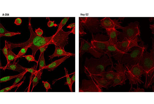


Jak/Stat Pathway Inhibitors Antibody Sampler Kit

 下载产品说明书 下载SDS
 用小程序,查商品更便捷


 收藏
收藏
 对比
对比 咨询
咨询
The Jak/Stat Pathway Inhibitors Antibody Sampler Kit provides an economical means to examine several inhibitors of Jak/Stat signaling, including PIAS1, PIAS3, PIAS4, SOCS1, SOCS2, and SOCS3. The kit contains enough primary antibody to perform two western blot experiments with each primary antibody.
参考图片
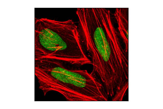
Confocal immunofluorescent analysis of A-204 (positive, left) and Hep G2 (low expression, right) cells using PIAS3 (D5F9) XP® Rabbit mAb (green). Actin filaments were labeled with DY-554 phalloidin (red).
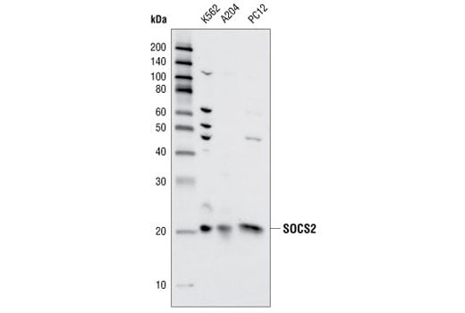
Western blot analysis of extracts from K562, A204 and PC12 cells using SOCS2 Antibody.
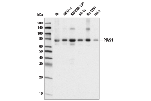
Western blot analysis of extracts from various cell lines using PIAS1 (D33A7) XP® Rabbit mAb.
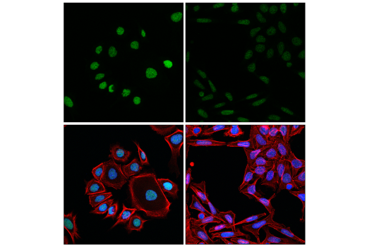
Confocal immunofluorescent analysis of AGS (left, high-expressing) and SW480 (right, low-expressing) using PIAS1 (D33A7) XP® Rabbit mAb (green), DyLight™ 650 Phalloidin #12956 (red), and DAPI #4083 (blue).
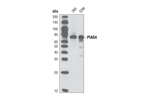
Western blot analysis of extracts from 293 and CCRF-CEM cells using PIAS4 (D2F12) Rabbit mAb.
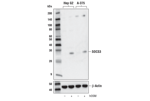
Western blot analysis of extracts from Hep G2 and A-375 cell lines, untreated (-) or treated with Human Oncostatin M #5367 (hOSM; 20 ng/ml, 1 hr; +), using SOCS3 (D6E1T) Rabbit mAb (upper) or β-Actin (D6A8) Rabbit mAb #8457 (lower).
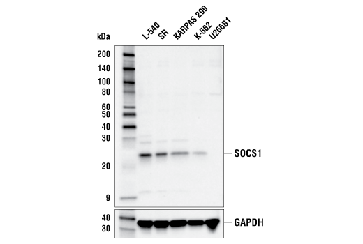
Western blot analysis of extracts from various cell lines using SOCS1 (E3Q4M) Rabbit mAb (upper) or GAPDH (D16H11) XP® Rabbit mAb #5174 (lower). The absence of signal for SOCS1 in U266B1 cells has been found to be caused by aberrant hypermethylation of the SOCS1 gene and confirms the specificity of the antibody for SOCS1.
After the primary antibody is bound to the target protein, a complex with HRP-linked secondary antibody is formed. The LumiGLO® is added and emits light during enzyme catalyzed decomposition.
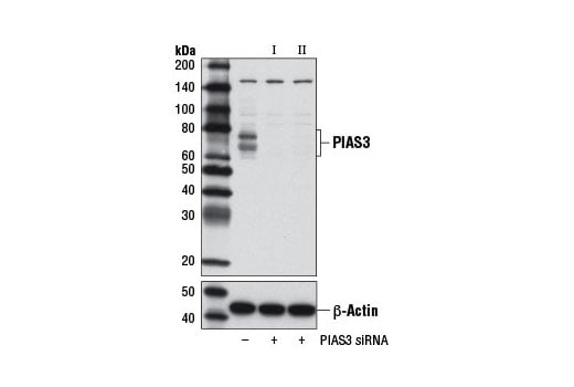
Western blot analysis of extracts from RD cells, transfected with 100 nM SignalSilence® Control siRNA (Unconjugated) #6568 (-), SignalSilence® PIAS3 siRNA I #9073 (+), or SignalSilence® PIAS3 siRNA II #9031 (+), using PIAS3 (D5F9) XP® Rabbit mAb (upper) or β-Actin (D6A8) Rabbit mAb #8457 (lower). The PIAS3 (D5F9) XP® Rabbit mAb confirms silencing of PIAS3 expression, while the β-Actin (D6A8) Rabbit mAb is used as a loading control.
Confocal imunofluorescent analysis of HeLa cells using PIAS1 (D33A7) XP® Rabbit mAb (green). Actin filaments have been labeled with DY-554 phalloidin (red).
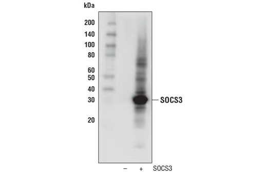
Western blot analysis of extracts from 293T cells, mock transfected (-) or transfected with a construct expressing full-length human SOCS3 (+), using SOCS3 (D6E1T) Rabbit mAb.
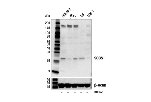
Western blot analysis of extracts from various cell lines, untreated or treated with mINFα (10ng/ml, 24 hr), using SOCS1 (E3Q4M) Rabbit mAb (upper) or β-Actin (D6A8) Rabbit mAb #8457 (lower).
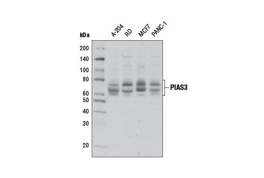
Western blot analysis of extracts from various cell lines using PIAS3 (D5F9) XP® Rabbit mAb.
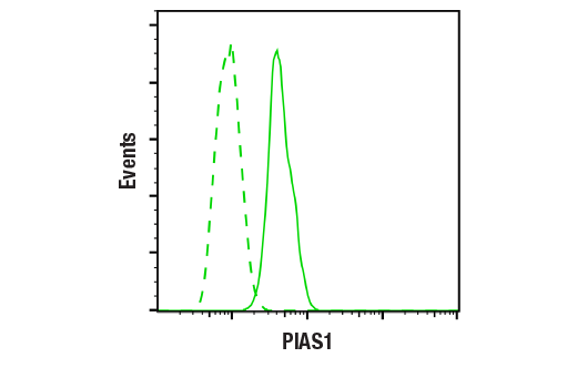
Flow cytometric analysis of KARPAS 299 cells using PIAS1 (D33A7) XP® Rabbit mAb (solid line) or concentration-matched Rabbit (DA1E) mAb IgG XP® Isotype control #3900 (dashed line). Anti-rabbit IgG (H+L), F(ab')2 Fragment (Alexa Fluor® 488 Conjugate) #4412 was used as a secondary antibody.
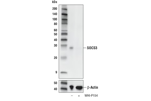
Western blot analysis of extracts from KARPAS-299 cells, untreated (-) or treated with the Jak3 inhibitor WHI-P154 (40 μM, overnight; +), using SOCS3 (D6E1T) Rabbit mAb (upper) or β-Actin (D6A8) Rabbit mAb (lower). KARPAS cell line source: Dr. Abraham Karpas at the University of Cambridge.
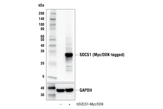
Western blot analysis of extracts from 293T cells, untransfected (-) or transfected with a construct expressing Myc/DDK-tagged full-length human SOCS1 protein (hSOCS1-Myc/DDK; +), using SOCS1 (E3Q4M) Rabbit mAb (upper) or GAPDH (D16H11) XP® Rabbit mAb #5174 (lower).
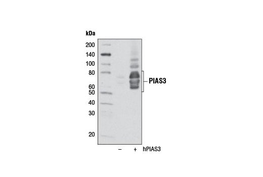
Western blot analysis of extracts from 293T cells, mock transfected (-) or transfected with a construct expressing full-length human PIAS3 (hPIAS3; +), using PIAS3 (D5F9) XP® Rabbit mAb.
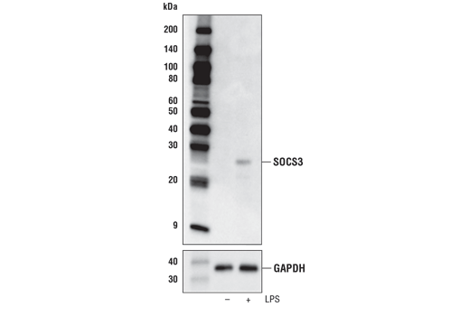
Western blot analysis of extracts from mouse bone marrow derived macrophage (mBMDM) cell extracts, untreated (-) or treated with Lipopolysaccharides #14011 (LPS; 50 ng/ml, 4 hr; +), using SOCS3 (D6E1T) Rabbit mAb (upper) or GAPDH (D16H11) XP® Rabbit mAb #5174 (lower).
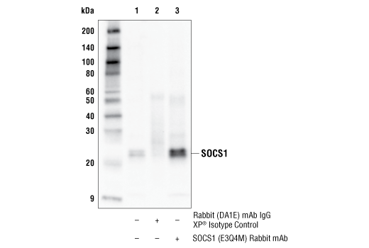
Immunoprecipitation of SOCS1 protein from L-540 cell extracts. Lane 1 is 10% input, lane 2 is Rabbit (DA1E) mAb IgG XP® Isotype Control #3900, and lane 3 is SOCS1 (E3Q4M) Rabbit mAb. Western blot analysis was performed using SOCS1 (E3Q4M) Rabbit mAb. Mouse Anti-rabbit IgG (Conformation Specific) (L27A9) mAb (HRP Conjugate) #5127 was used as a secondary antibody.