


 全部商品分类
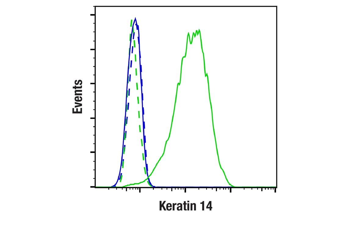


 用小程序,查商品更便捷


Monoclonal antibody is produced by immunizing animals with a synthetic peptide corresponding to residues near the carboxy terminus of human keratin 14 protein.

Product Usage Information
This product is the carrier free version of product #74956. All data were generated using the same antibody clone in the standard formulation which contains BSA and glycerol.
This formulation is ideal for use with technologies requiring specialized or custom antibody labeling, including fluorophores, metals, lanthanides, and oligonucleotides. It is not recommended for ChIP, ChIP-seq, CUT&RUN or CUT&Tag assays. If you require a carrier free formulation for chromatin profiling, please contact us. Optimal dilutions/concentrations should be determined by the end user.
BSA and Azide Free antibodies are quality control tested by size exclusion chromatography (SEC) to determine antibody integrity.
Specificity/Sensitivity
Species Reactivity:
Human, Mouse
Store at -20°C. This product will freeze at -20°C so it is recommended to aliquot into single-use vials to avoid multiple freeze/thaw cycles. A slight precipitate may be present and can be dissolved by gently vortexing. This will not interfere with antibody performance.
参考图片
Flow cytometric analysis of PANC-1 cells (blue, negative) and A-431 cells (green, positive) using Keratin 14 (E7W6V) Rabbit mAb (solid lines) or concentration-matched Rabbit (DA1E) mAb IgG XP® Isotype Control #3900 (dashed lines). Anti-rabbit IgG (H+L), F(ab')2 Fragment (Alexa Fluor® 488 Conjugate) #4412 was used as a secondary antibody. Data were generated using the standard formulation of this product.
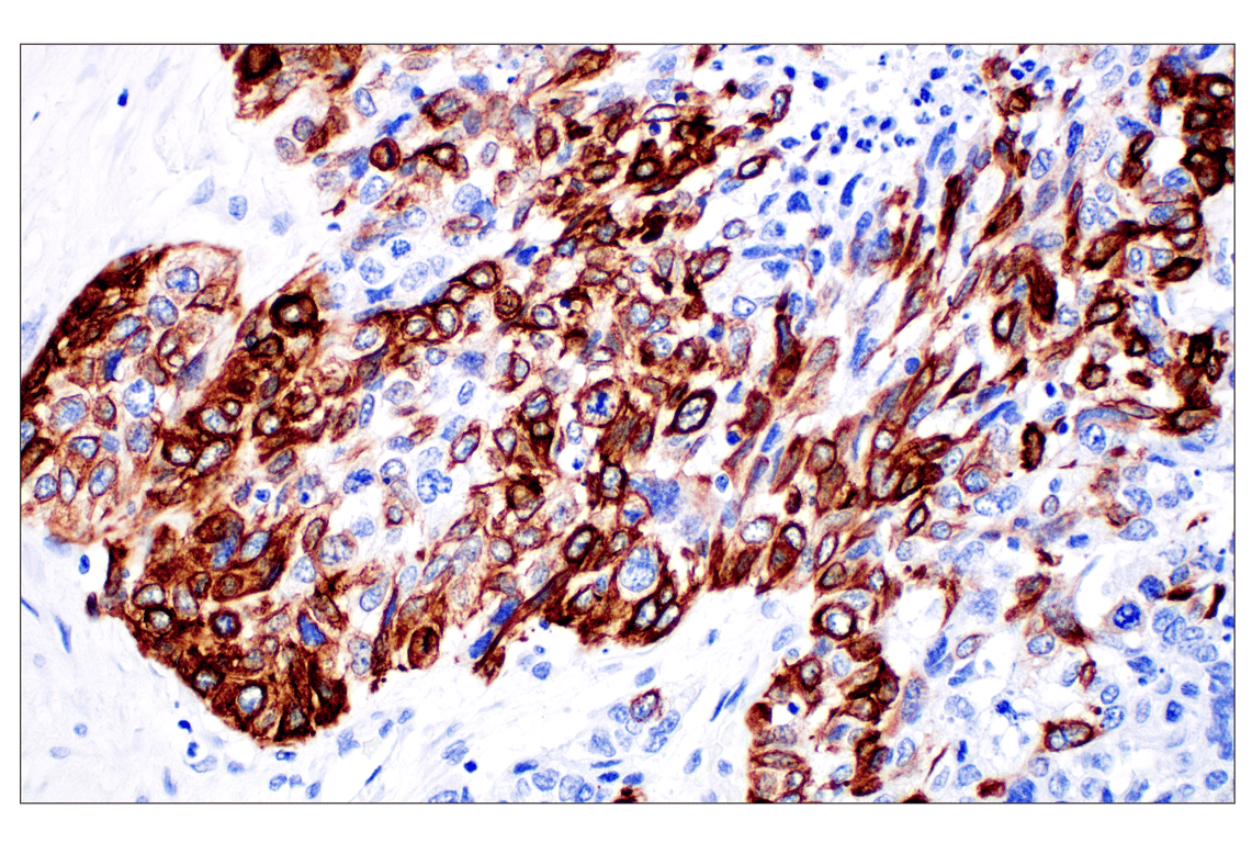
Immunohistochemical analysis of paraffin-embedded human urothelial carcinoma using Keratin 14 (E7W6V) Rabbit mAb. Data were generated using the standard formulation of this product.
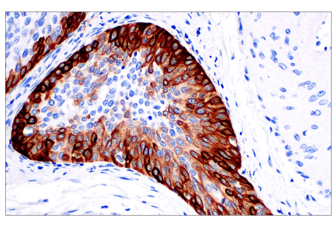
Immunohistochemical analysis of paraffin-embedded human squamous cell carcinoma of the cervix using Keratin 14 (E7W6V) Rabbit mAb. Data were generated using the standard formulation of this product.
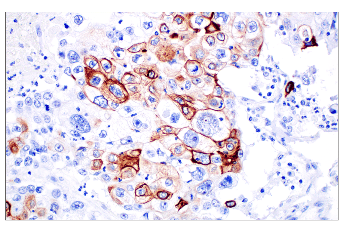
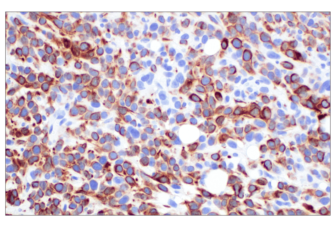
Immunohistochemical analysis of paraffin-embedded human hepatocellular carcinoma using Keratin 14 (E7W6V) Rabbit mAb. Data were generated using the standard formulation of this product.
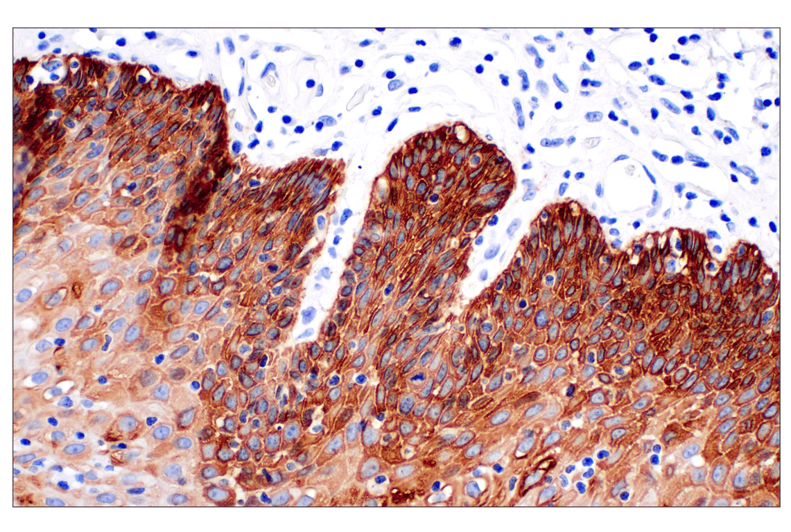
Immunohistochemical analysis of paraffin-embedded normal human esophagus using Keratin 14 (E7W6V) Rabbit mAb. Data were generated using the standard formulation of this product.
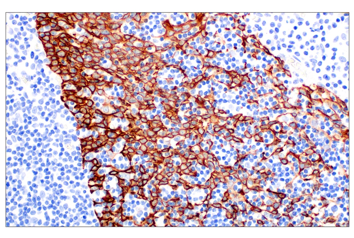
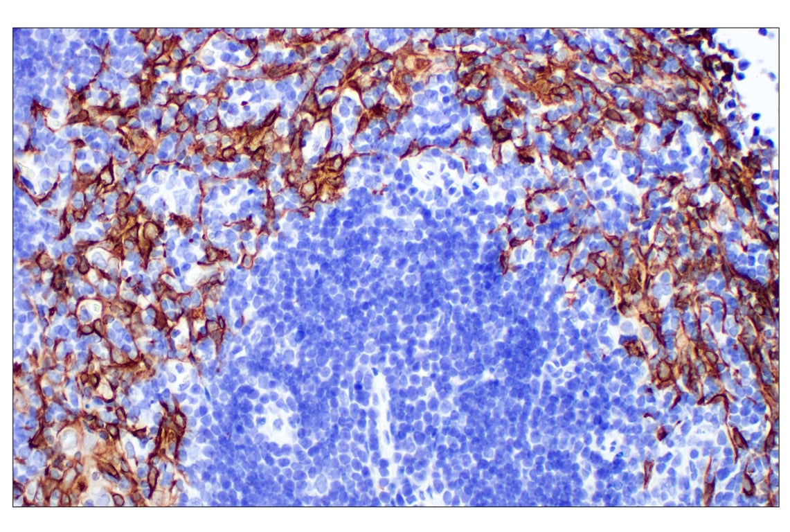
Immunohistochemical analysis of paraffin-embedded human follicular hyperplasia of the tonsil using Keratin 14 (E7W6V) Rabbit mAb. Data were generated using the standard formulation of this product.
Immunohistochemical analysis of paraffin-embedded 4T1 syngeneic tumor using Keratin 14 (E7W6V) Rabbit mAb. Data were generated using the standard formulation of this product.
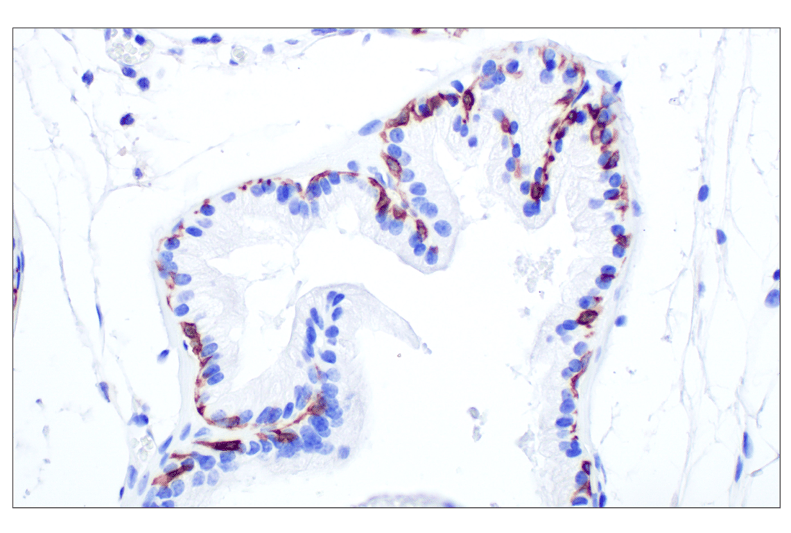
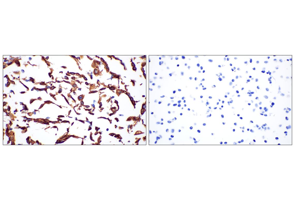
Immunohistochemical analysis of paraffin-embedded mouse prostate using Keratin 14 (E7W6V) Rabbit mAb. Data were generated using the standard formulation of this product.
Immunohistochemical analysis of paraffin-embedded mouse thymus using Keratin 14 (E7W6V) Rabbit mAb. Data were generated using the standard formulation of this product.
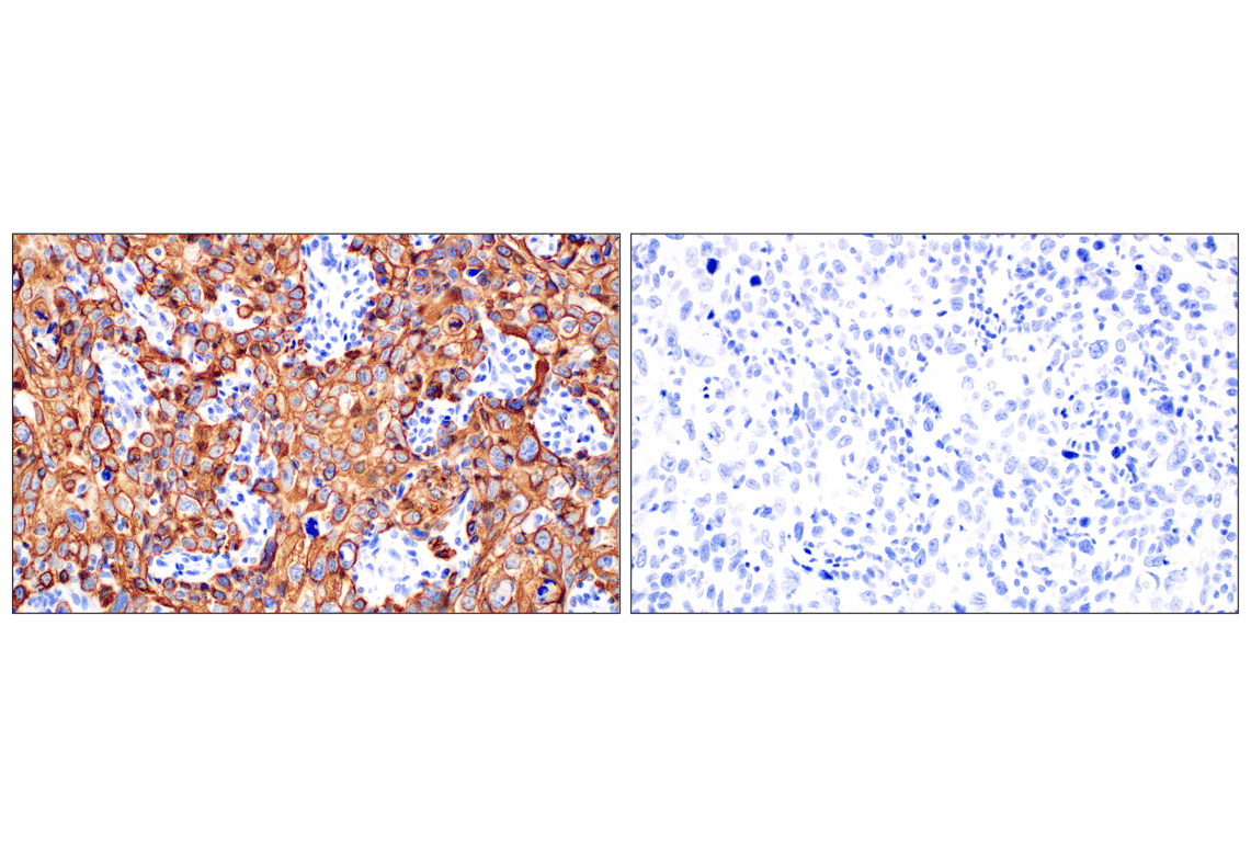
Immunohistochemical analysis of paraffin-embedded human oropharyngeal squamous cell carcinoma using Keratin 14 (E7W6V) Rabbit mAb (left) compared to concentration-matched Rabbit (DA1E) mAb IgG XP® Isotype Control #3900 (right). Data were generated using the standard formulation of this product.
Immunohistochemical analysis of paraffin-embedded A-431 cell pellet (left, positive) or A-673 cell pellet (right, negative) using Keratin 14 (E7W6V) Rabbit mAb. Data were generated using the standard formulation of this product.
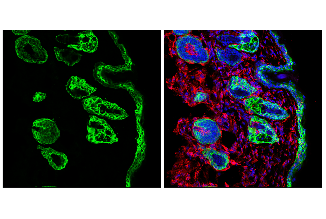
Confocal immunofluorescent analysis of fixed frozen mouse skin labeled with Keratin 14 (E7W6V) Rabbit mAb (left, green). Free secondary binding sites were then blocked with Rabbit (DA1E) mAb IgG XP® Isotype Control #3900 prior to co-labeling with Vimentin (D21H3) XP® Rabbit mAb (Alexa Fluor® 555 Conjugate) #9855 (right, red) and ProLong Gold Antifade Reagent with DAPI #8961 (right, blue). Data were generated using the standard formulation of this product.
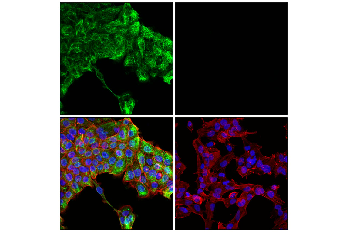
Confocal immunofluorescent analysis of A-431 cells (left, positive) and A-673 cells (right, negative) using Keratin 14 (E7W6V) Rabbit mAb (green), DyLight 554 Phalloidin #13054 (red), and DAPI #4083 (blue). Data were generated using the standard formulation of this product.