


 全部商品分类
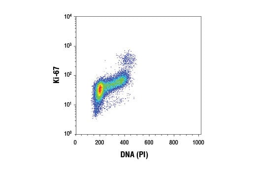


Ki-67 (D3B5) Rabbit mAb


 下载产品说明书 下载COA 下载SDS
 用小程序,查商品更便捷


 收藏
收藏
 对比
对比 咨询
咨询
Monoclonal antibody is produced by immunizing animals with a recombinant protein fragment corresponding to residues near the central region of human Ki-67 protein.
Product Usage Information
| Application | Dilution |
|---|---|
| Immunofluorescence (Frozen) | 1:400 - 1:800 |
| Immunofluorescence (Immunocytochemistry) | 1:400 - 1:800 |
| Flow Cytometry (Fixed/Permeabilized) | 1:400 - 1:1600 |
Specificity/Sensitivity
Species Reactivity:
Human, Mouse, Rat
Supplied in 10 mM sodium HEPES (pH 7.5), 150 mM NaCl, 100 µg/ml BSA, 50% glycerol and less than 0.02% sodium azide. Store at –20°C. Do not aliquot the antibody.
For a carrier free (BSA and azide free) version of this product see product #34330.
参考图片
Flow cytometric analysis of Jurkat cells using Ki-67 (D3B5) Rabbit mAb and Propidium Iodide (PI)/RNase Staining Solution #4087. Anti-rabbit IgG (H+L), F(ab')2 Fragment (Alexa Fluor® 488 Conjugate) #4412 was used as a secondary antibody.
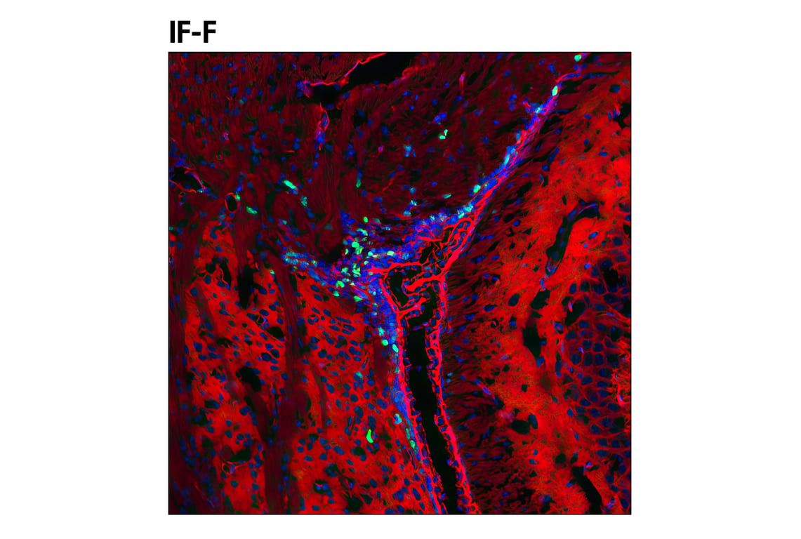
Confocal immunofluorescent analysis of the ventricular zone in P21 mouse brain using Ki-67 (D3B5) Rabbit mAb (green). Actin filaments were labeled with DyLight™ 554 phalloidin #13054 (red). Blue pseudocolor = DRAQ5® #4084 (fluorescent DNA dye).
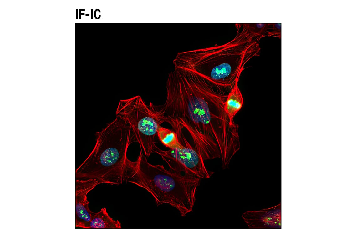
Confocal immunofluorescent analysis of HeLa cells using Ki-67 (D3B5) Rabbit mAb (green). Actin filaments were labeled with DY-554 phalloidin (red). Blue pseudocolor = DRAQ5® #4084 (fluorescent DNA dye).