


 全部商品分类


 用小程序,查商品更便捷


Monoclonal antibody is produced by immunizing animals with a synthetic peptide corresponding to residues near the amino terminus of human Ki-67 protein.
Product Usage Information
| Application | Dilution |
|---|---|
| Immunohistochemistry (Paraffin) | 1:800 - 1:3200 |
| Immunofluorescence (Immunocytochemistry) | 1:1600 - 1:6400 |
| Flow Cytometry (Fixed/Permeabilized) | 1:100 - 1:400 |
Specificity/Sensitivity
Species Reactivity:
Human, Monkey
Supplied in 10 mM sodium HEPES (pH 7.5), 150 mM NaCl, 100 µg/ml BSA, 50% glycerol and less than 0.02% sodium azide. Store at –20°C. Do not aliquot the antibody.
For a carrier-free (BSA and azide free) version of this product see product #62548.
参考图片
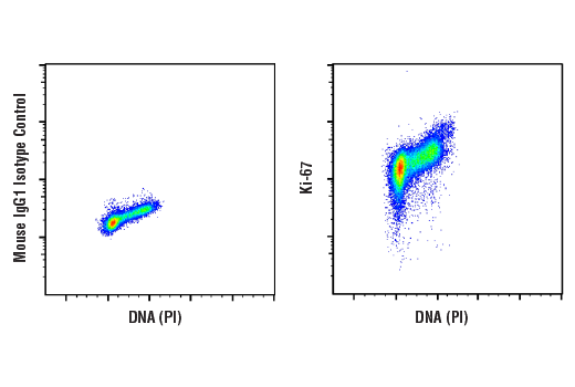
Flow cytometric analysis of Jurkat cells using Ki-67 (8D5) Mouse mAb (right) and Propidium Iodide (PI)/RNase Staining Solution #4087, compared to concentration-matched Mouse (G3A1) mAb IgG1 Isotype Control #5415 (left). Anti-mouse IgG (H+L), F(ab')2 Fragment (Alexa Fluor® 488 Conjugate) #4408 was used as a secondary antibody.
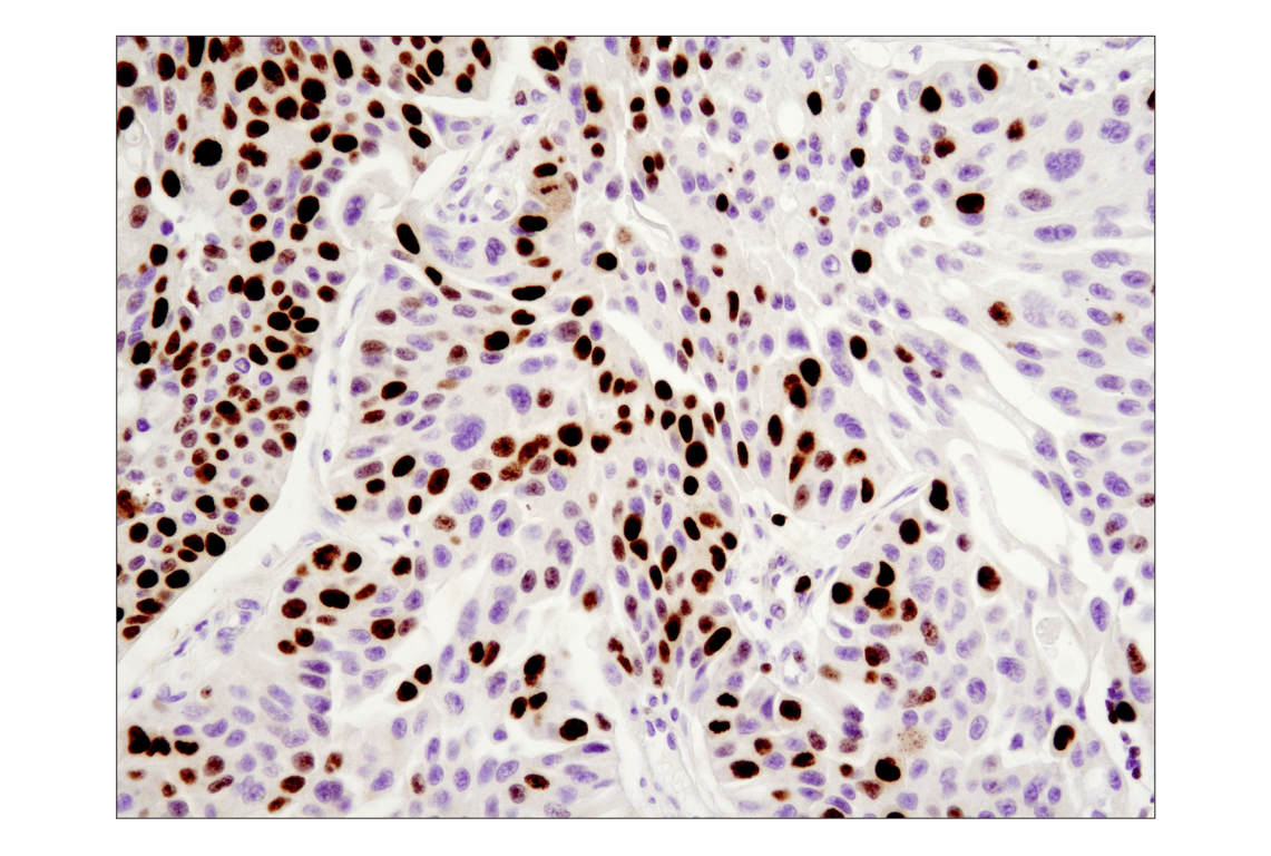
Immunohistochemical analysis of paraffin-embedded human breast carcinoma using Ki-67 (8D5) Mouse mAb.
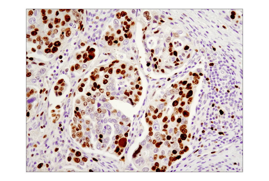
Immunohistochemical analysis of paraffin-embedded human colon carcinoma using Ki-67 (8D5) Mouse mAb.
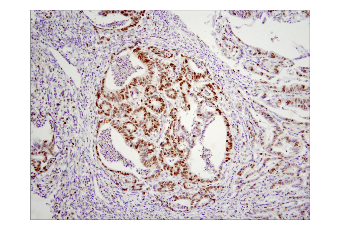
Immunohistochemical analysis of paraffin-embedded human ovarian serous adenocarcinoma using Ki-67 (8D5) Mouse mAb.
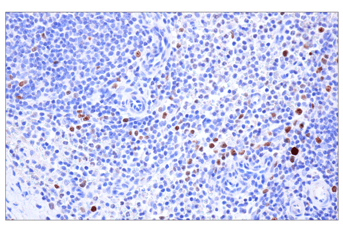
Immunohistochemical analysis of paraffin-embedded rhesus monkey spleen using Ki-67 (8D5) Mouse mAb.
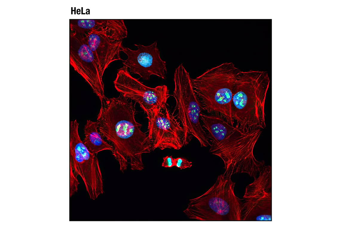
Confocal immunofluorescent analysis of HeLa cells using Ki-67 (8D5) Mouse mAb (green). Actin filaments were labeled with DY-554 phalloidin (red). Blue pseudocolor = DRAQ5® #4084 (fluorescent DNA dye).