


 全部商品分类
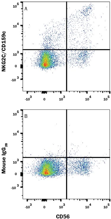


 下载产品说明书 下载SDS
 用小程序,查商品更便捷


 收藏
收藏
 对比
对比 咨询
咨询
Scientific Data
 View Larger
Detection of NKG2C/CD159c in Human Blood Lymphocytes by Flow Cytometry. Human peripheral blood lymphocytes were stained with Mouse Anti-Human NCAM-1/CD56 APC-conjugated Monoclonal Antibody (Catalog # FAB2408A) and either (A) Mouse Anti-Human NKG2C/CD159c Monoclonal Antibody (Catalog # MAB1381) or (B) Mouse IgG2BFlow Cytometry Isotype Control (Catalog # MAB0041) followed by Phycoerythrin-conjugated Anti-Mouse IgG Secondary Antibody (Catalog # F0102B). View our protocol for Staining Membrane-associated Proteins.
Human NKG2C/CD159c Antibody Summary
cross-reactivity with the human NKG2A/CD94 heterodimer or with the human CD94 homodimer is detected.
Applications
Please Note: Optimal dilutions should be determined by each laboratory for each application. General Protocols are available in the Technical Information section on our website.
Background: NKG2C/CD159c
Human NKG2C (NK cell Group 2 isoform C; Killer cell lectin-like receptor subfamily C, member 2) is a member of the C-type lectin-like superfamily of proteins. Natural killer (NK) receptors are expressed in both NK cells and cytotoxic CD8+ T cells and have both activating and inhibitory members (1-3). Regulation of the balance between the activating and inhibitory receptors is important and lack of such regulation has been implicated in autoimmunity (4). The NKG2 family includes seven receptors: NKG2A, -B, -C, -D, -E, -F, and -H, which is the longer isoform of NKG2E. Except for NKG2D and NKG2F, the NKG2 family members form heterodimers with CD94 (5, 6). NKG2C interacts with the adapter molecule DAP12 and acts as activating receptor when heterodimerized with CD94 (7). Human NKG2C is synthesized as a 231 amino acid (aa) protein that includes a 70 aa cytoplasmic domain, a 23 aa transmembrane segment, and a 138 aa extracellular domain (ECD). Within the ECD, human NKG2C shares 40% sequence identity with mouse NKG2C. NKG2C-CD94 heterodimers bind to the widely expressed nonclassical MHC-I molecule, HLA-E (Qa-1b in mouse), which presents a peptide derived from the signal peptide of classical MHC-I molecules (8, 9). Triggering the NKG2C-CD94 complex may activate the cytolytic activity and cytokine production of NK and CD8+ T cells (8, 10). Human cytomegalovirus (HCMV) infection promotes the differentiation and expansion of NKG2C+ NK cell subsets, possibly involving a cognate interaction of CD94/NKG2C with ligand(s) displayed by HCMV‑infected cells (11, 12). MAB1381 (clone 134522) displays potent agonistic activity and also blocks the binding of the NKG2C/CD94 heterodimer to HLA-E tetramers (13-15).
- Orbelyan, G.A. et al. (2014) J. Immunol. 193:610.
- Tassi I. et al. (2006) Immnunol Rev. 214:92.
- Lanier, L.L. (2008) Nat. Immunol. 9:495.
- Schleinitz, N. et al. (2010) Immunology. 174:2878.
- Lopez-Botet, M. et al. (2000) Hum. Immunol. 61:7.
- Braud, V.M. et al. (1998) Nature. 391:795.
- Lanier, L.L. (1998) Immunity 8:693.
- Vance, R.E. et al. (1999) J Exp Med 190:1801.
- Kaiser B.K. et al. (2005) J Immunol 174:2878.
- Bellón T. et al. (1999) J Immunol 162:3996.
- Pupuleku A. et al. (2017) Front Immunol. 8:1317.
- Hammer Q. et al. (2018) Nat Immunol. 19:453.
- Alici, E. et al. (2008) Blood 111:3155.
- Coupel, S. et al. (2007) Blood 109:2806.
- Fausther-Bovendo, H. et al. (2008) AIDS 22:217.
Preparation and Storage
- 12 months from date of receipt, -20 to -70 °C as supplied.
- 1 month, 2 to 8 °C under sterile conditions after reconstitution.
- 6 months, -20 to -70 °C under sterile conditions after reconstitution.
参考图片
Detection of NKG2C/CD159c in Human Blood Lymphocytes by Flow Cytometry. Human peripheral blood lymphocytes were stained with Mouse Anti-Human NCAM‑1/CD56 APC‑conjugated Monoclonal Antibody (Catalog # FAB2408A) and either (A) Mouse Anti-Human NKG2C/CD159c Monoclonal Antibody (Catalog # MAB1381) or (B) Mouse IgG2B Flow Cytometry Isotype Control (Catalog # MAB0041) followed by Phycoerythrin-conjugated Anti-Mouse IgG Secondary Antibody (Catalog # F0102B). View our protocol for Staining Membrane-associated Proteins.